. 2008 Dec 19;283(51):35756-62.
doi: 10.1074/jbc.M807125200. Epub 2008 Sep 29.

Human carbonyl reductase 1 is an S-nitrosoglutathione reductase

Affiliations

Human carbonyl reductase 1 is an S-nitrosoglutathione reductase

Raynard L Bateman et al. J Biol Chem. .

Abstract

Human carbonyl reductase 1 (hCBR1) is an NADPH-dependent short chain dehydrogenase/reductase with broad substrate specificity and is thought to be responsible for the in vivo reduction of quinones, prostaglandins, and other carbonyl-containing compounds including xenobiotics. In addition, hCBR1 possesses a glutathione binding site that allows for increased affinity toward GSH-conjugated molecules. It has been suggested that the GSH-binding site is near the active site; however, no structures with GSH or GSH conjugates have been reported. We have solved the x-ray crystal structures of hCBR1 and a substrate mimic in complex with GSH and the catalytically inert GSH conjugate hydroxymethylglutathione (HMGSH). The structures reveal the GSH-binding site and provide insight into the affinity determinants for GSH-conjugated substrates. We further demonstrate that the structural isostere of HMGSH, S-nitrosoglutathione, is an ideal hCBR1 substrate (Km = 30 microm, kcat = 450 min(-1)) with kinetic constants comparable with the best known hCBR1 substrates. Furthermore, we demonstrate that hCBR1 dependent GSNO reduction occurs in A549 lung adenocarcinoma cell lysates and suggest that hCBR1 may be involved in regulation of tissue levels of GSNO.


Figures
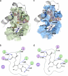
FIGURE 1.
FIGURE 1.
The glutathione binding site of CBR1. a, electron density maps of hCBR1·OH-PP·NADP·GSH. b, binding mode of hCBR1·OH-PP·NADP·GSH. c, electron density maps of hCBR1·OH-PP·NADP·HMGSH. d, binding mode of hCBR1·OH-PP·NADP·HMGSH. For a and c, the 2Fo - Fc maps are contoured in brown at a level of 1σ. NADP, OH-PP, and either GSH or HMGSH (yellow, red, blue, and white) are shown occupying the CBR1 active site. For b and d, hydrogen bonding interactions and residues within a distance of 4.0 Å from the ligand are shown. Nonpolar residues (green), polar residues (purple), and charged residues (purple/blue) are indicated. The distances are given (Å).
FIGURE 2.
FIGURE 2.
Comparison of hCBR1·NADP (a) and hCBR1·NADP·GSH (b) structures. Either a halogen (a) or the C226 Sγ thiol/thiolate (b) is stabilized by H-bonding interactions with main chain amines of Ile140 and Gly228. The anion-binding site is also at the N terminus of an α-helix.
FIGURE 3.
FIGURE 3.
Overlay of hCBR1·OH-PP·NADP (Protein Data Bank code 1WMA, pink), hCBR1·OH-PP·NADP·GSH (Protein Data Bank code 3BHJ, olive), hCBR1·OH-PP·NADP·HMGSH (Protein Data Bank code 3BHM, blue), and hCBR1·NADP (Protein Data Bank code 3BHI, yellow). NADP, HMGSH, and OH-PP are indicated. The flexible regions Lys95–Pro102 and Met234–Lys238 are shown.
SCHEME 1
SCHEME 1
FIGURE 4.
FIGURE 4.
NADH- and NADPH-dependent GSNO reductase activity in A549 cell lysates with and without the presence of the selective hCBR1 inhibitor, OH-PP-Me (details under “Experimental Procedures”).

References

    1. Forrest, G. L., and Gonzalez, B. (2000) Chem. Biol. Interact 129 21-40 - PubMed
    1. Doorn, J. A., Maser, E., Blum, A., Claffey, D. J., and Petersen, D. R. (2004) Biochemistry 43 13106-13114 - PubMed
    1. Tanaka, M., Bateman, R., Rauh, D., Vaisberg, E., Ramachandani, S., Zhang, C., Hansen, K. C., Burlingame, A. L., Trautman, J. K., Shokat, K. M., and Adams, C. L. (2005) PLoS Biol. 3 764-776 - PMC - PubMed
    1. Tinguely, J. N., and Wermuth, B. (1999) Eur. J. Biochem. 260 9-14 - PubMed
    1. Bateman, R., Rauh, D., and Shokat, K. M. (2007) Org. Biomol. Chem. 5 3363-3367 - PMC - PubMed

Publication types

MeSH terms

Substances