. 2020 Feb 7;295(6):1646-1657.
doi: 10.1074/jbc.RA119.011758. Epub 2020 Jan 6.

The bacterial deubiquitinase Ceg23 regulates the association of Lys-63-linked polyubiquitin molecules on the Legionella phagosome

Affiliations

The bacterial deubiquitinase Ceg23 regulates the association of Lys-63-linked polyubiquitin molecules on the Legionella phagosome

Kelong Ma et al. J Biol Chem. .

Abstract

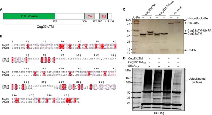
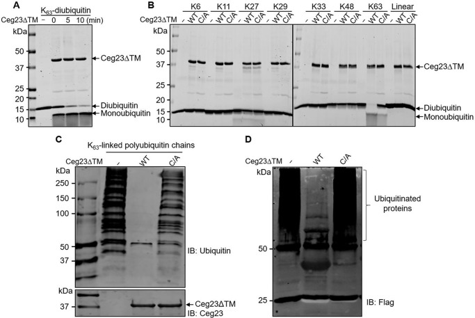
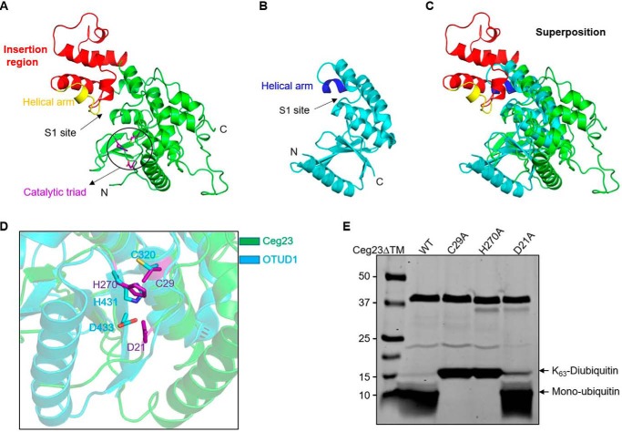
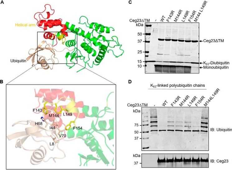
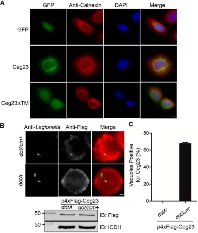
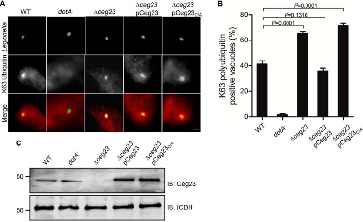
Legionella pneumophila is the causative agent of the lung malady Legionnaires' disease, it modulates host function to create a niche termed the Legionella-containing vacuole (LCV) that permits intracellular L. pneumophila replication. One important aspect of such modulation is the co-option of the host ubiquitin network with a panel of effector proteins. Here, using recombinantly expressed and purified proteins, analytic ultracentrifugation, structural analysis, and computational modeling, along with deubiquitinase (DUB), and bacterial infection assays, we found that the bacterial defective in organelle trafficking/intracellular multiplication effector Ceg23 is a member of the ovarian tumor (OTU) DUB family. We found that Ceg23 displays high specificity toward Lys-63-linked polyubiquitin chains and is localized on the LCV, where it removes ubiquitin moieties from proteins ubiquitinated by the Lys-63-chain type. Analysis of the crystal structure of a Ceg23 variant lacking two putative transmembrane domains at 2.80 Å resolution revealed that despite very limited homology to established members of the OTU family at the primary sequence level, Ceg23 harbors a catalytic motif resembling those associated with typical OTU-type DUBs. ceg23 deletion increased the association of Lys-63-linked polyubiquitin with the bacterial phagosome, indicating that Ceg23 regulates Lys-63-linked ubiquitin signaling on the LCV. In summary, our findings indicate that Ceg23 contributes to the regulation of the association of Lys-63 type polyubiquitin with the Legionella phagosome. Future identification of host substrates targeted by Ceg23 could clarify the roles of these polyubiquitin chains in the intracellular life cycle of L. pneumophila and Ceg23's role in bacterial virulence.

Keywords: Legionella pneumophila; bacteria; bacterial pathogenesis; deubiquitylation (deubiquitination); effector protein; ovarian-tumor family deubiquitinases; polyubiquitin chain; structural biology; type IV secretion system.


Conflict of interest statement

The authors declare that they have no conflicts of interest with the contents of this article

Figures

Figure 1.
Figure 1.
Ceg23 shows DUB activity in vitro. A, a diagram of the predicted domain structure of Ceg23. B, sequence alignment of Ceg23 with OTUB1 (NCBI reference sequence NP_060140.2), a member of the eukaryotic OTU protein DUB superfamily. Alignment was generated by Clustal Omega and ESPript 3.0. Residues with the red background indicate identical sites, and residues highlighted by blue boxes represent conserved sites. Asterisks indicate the cysteine and histidine residues potentially critical for catalysis. C, Ceg23ΔTM but not Ceg23ΔTMC29A reacted with the suicide probe Ub-PA. LotA, a Legionella OTU-like DUB, was used as a positive control. D, Ceg23ΔTM removes ubiquitin chains from modified proteins. Polyubiquitinated proteins immunoprecipitated using agarose beads coated with the anti-FLAG antibody from HEK293T cells transfected to express 4× FLAG–Ub were incubated with Ceg23ΔTM or Ceg23ΔTMC29A. Ubiquitin signals were detected by the anti-FLAG antibody. The established DUB SdeADUB was included as a positive control. The results in C and D show one representative from three independent experiments.
Figure 2.
Figure 2.
Ceg23 specifically cleaves Lys-63–linked ubiquitin chains. A, time-dependent cleavage of Lys-63–linked diubiquitin by Ceg23ΔTM. 1 μm of Ceg23ΔTM was incubated with 1 μm of diubiquitin at 37 °C for the indicated time. B, Ceg23ΔTM induced cleavage of diubiquitins linked by the primary methionine and each of the seven lysine residues. 1 μm of Ceg23ΔTM was incubated with 1 μm of diubiquitin at 37 °C for 2 h, and DUB activity was indicated by the release of free ubiquitin. C and D, disassembly of Lys-63–linked polyubiquitin chains by Ceg23ΔTM. Polyubiquitin chains assembled by Lys-63 were either synthesized by an in vitro reaction containing ubiquitin, E1, UBE2V2, and UBE2N (C) or isolated from cells transfected with pCMV–4× FLAG–Ub63K (a ubiquitin mutant that harbors only Lys-63) (D). After incubation with Ceg23ΔTM, polyubiquitin was probed by antibodies specific for ubiquitin or for the FLAG tag. The results in A–D show one representative from three independent experiments. IB, immunoblot.
Figure 3.
Figure 3.
Structural features of Ceg23ΔTM. A, structure of Ceg23ΔTM in ribbon representations. The insertion region is colored in red, and the helical arm for ubiquitin binding is highlighted in yellow. Residues participating in the formation of the catalytic site shown with purple sticks are highlighted by a circle. B, crystal structure of the human OTUD1 (PDB code 4BOP), one of the OTU family DUBs. The ubiquitin-contacting helical arm is colored in blue. C, superposition of Ceg23ΔTM (colored as in A) and OTUD1 (colored as in B). D, zoom-in view of the aligned active site regions of Ceg23ΔTM and OTUD1. E, cleavage of Lys-63–linked diubiquitin by C29A, H270A, and D21A mutants of Ceg23ΔTM. 1 μm of Ceg23ΔTM was incubated with 1 μm of Lys-63–linked diubiquitin at 37 °C for 5 min. Note that mutation in Cys-29 or H270A almost completely abolished its activity, whereas mutation in Asp-21 only slightly affects its ability to cleave Lys-63–linked diubiquitin. E is one representative from three independent experiments.
Figure 4.
Figure 4.
Docking of ubiquitin into structure of Ceg23ΔTM. A, the predicted ubiquitin recognition mode of Ceg23ΔTM. B, ubiquitin inserted into a hydrophobic cavity of Ceg23ΔTM formed by residues Phe-143, Met-144, Leu-149, and Phe-154. C and D, cleavage of Lys-63–linked diubiquitin (C) or polyubiquitin chains (D) by F143R, M144R, L149R, F154R, and M144R/L149R mutants of Ceg23ΔTM. The reactions were performed at 37 °C for 5 min. Note that single mutation of Phe-143, Met-144, Leu-149, or Phe-154 only slightly reduced its activity, whereas simultaneous mutations in Met-144 and Leu-149 resulted in an almost complete loss of its DUB activity. C and D are one representative from three independent experiments.
Figure 5.
Figure 5.
Cellular localization of Ceg23. A, Ceg23 localized to the ER in a manner that requires the two predicted transmembrane domains. HeLa cells were transfected with plasmids that direct the expression of GFP–Ceg23 or GFP–Ceg23ΔTM. The ER was labeled by immunostaining with antibodies specific for the ER-resident protein calnexin. Bar, 5 μm. B, the association of Ceg23 with the LCV. U937 cells infected with the indicated L. pneumophila strains expressing 4× FLAG–Ceg23 were sequentially immunostained with antibodies against L. pneumophila and FLAG. Images were obtained by a fluorescence microscope. The lower panel indicated that 4× FLAG–Ceg23 was similarly expressed in dotA and dot/icm+ strains. C, quantification of LCVs positively stained for 4× FLAG–Ceg23 shown in B. The results in B and C are from one representative experiment done in triplicate from three independent experiments; at least 100 vacuoles were counted in each sample. Bar, 5 μm. The error bars represent S.E. (n = 3). DAPI, 4′,6′-diamino-2-phenylindole; IB, immunoblot.
Figure 6.
Figure 6.
Ceg23 down-regulates the association of Lys-63–linked polyubiquitins with the LCV. A, BMDMs were challenged with the indicated L. pneumophila strains for 2 h. The cells were immunostained with antibodies against L. pneumophila and Lys-63–linked polyubiquitin, respectively. Representative images were taken by a fluorescence microscope. Bar, 5 μm. B, quantification of phagosomes associated with Lys-63–linked polyubiquitin. The data in B show one representative experiment done in triplicate from three independent experiments. At least 100 vacuoles were counted in each sample. The error bars represent S.E. (n = 3). The p values were calculated by unpaired two-tailed Student's t tests, and p < 0.05 was considered as significant difference. C, expression of Ceg23 in the L. pneumophila strains used for infection. The metabolic enzyme isocitrate dehydrogenase (ICDH) was probed as a loading control. C shows one representative from three independent experiments. IB, immunoblot.

References

    1. Hershko A., and Ciechanover A. (1998) The ubiquitin system. Annu. Rev. Biochem. 67, 425–479 10.1146/annurev.biochem.67.1.425 - DOI - PubMed
    1. Pisano A., Albano F., Vecchio E., Renna M., Scala G., Quinto I., and Fiume G. (2018) Revisiting bacterial ubiquitin ligase effectors: weapons for host exploitation. Int. J. Mol. Sci. 19, E3576 10.3390/ijms19113576 - DOI - PMC - PubMed
    1. Komander D., and Rape M. (2012) The ubiquitin code. Annu. Rev. Biochem. 81, 203–229 10.1146/annurev-biochem-060310-170328 - DOI - PubMed
    1. Yau R., and Rape M. (2016) The increasing complexity of the ubiquitin code. Nat. Cell Biol. 18, 579–586 10.1038/ncb3358 - DOI - PubMed
    1. Walczak H., Iwai K., and Dikic I. (2012) Generation and physiological roles of linear ubiquitin chains. BMC Biol. 10, 23 10.1186/1741-7007-10-23 - DOI - PMC - PubMed

Publication types

LinkOut - more resources