. 2008 Jul 1;105(26):9065-9.
doi: 10.1073/pnas.0800502105. Epub 2008 Jun 11.

Structure of coronavirus hemagglutinin-esterase offers insight into corona and influenza virus evolution

Affiliations

Structure of coronavirus hemagglutinin-esterase offers insight into corona and influenza virus evolution

Qinghong Zeng et al. Proc Natl Acad Sci U S A. .

Abstract

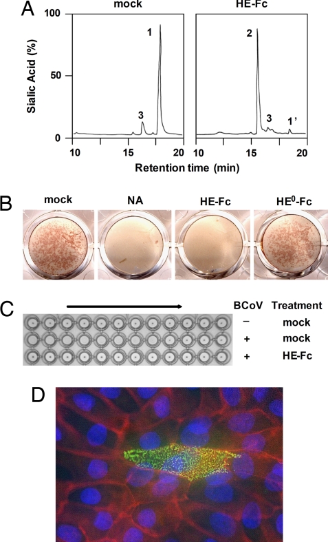
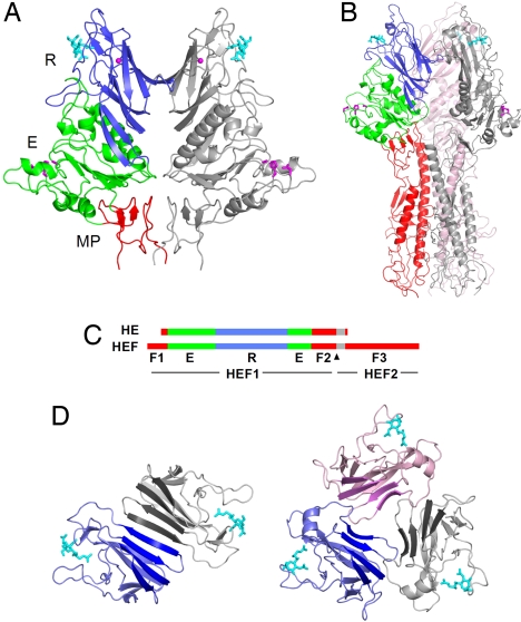
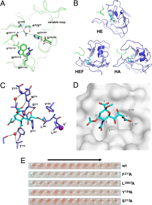
The hemagglutinin-esterases (HEs) are a family of viral envelope glycoproteins that mediate reversible attachment to O-acetylated sialic acids by acting both as lectins and as receptor-destroying enzymes (RDEs). Related HEs occur in influenza C, toro-, and coronaviruses, apparently as a result of relatively recent lateral gene transfer events. Here, we report the crystal structure of a coronavirus (CoV) HE in complex with its receptor. We show that CoV HE arose from an influenza C-like HE fusion protein (HEF). In the process, HE was transformed from a trimer into a dimer, whereas remnants of the fusion domain were adapted to establish novel monomer-monomer contacts. Whereas the structural design of the RDE-acetylesterase domain remained unaltered, the HE receptor-binding domain underwent remodeling to such extent that the ligand is now bound in opposite orientation. This is surprising, because the architecture of the HEF site was preserved in influenza A HA over a much larger evolutionary distance, a switch in receptor specificity and extensive antigenic variation notwithstanding. Apparently, HA and HEF are under more stringent selective constraints than HE, limiting their exploration of alternative binding-site topologies. We attribute the plasticity of the CoV HE receptor-binding site to evolutionary flexibility conferred by functional redundancy between HE and its companion spike protein S. Our findings offer unique insights into the structural and functional consequences of independent protein evolution after interviral gene exchange and open potential avenues to broad-spectrum antiviral drug design.


Conflict of interest statement

The authors declare no conflict of interest.

Figures

Fig. 1.
Fig. 1.
BCoV HE-Fc chimeras retain esterase and lectin activity. (A) HE-Fc displays sialate-9-O-acetylesterase activity. Purified HE-Fc was assayed for substrate specificity as described (16) with the synthetic di-O-acetylated sialic acid analogue αNeu4,5,9Ac32Me as substrate. Graphs show total ion current gas chromatograms. Sialic acids were identified by mass spectrometry: αNeu4,5,9Ac32Me (peak 1), αNeu4,5Ac22Me (peak 2); peaks 1′ and 3 represent nonsialic acid compounds. (B) HE-Fc, but not esterase-deficient HE0-Fc, destroys BCoV receptors on the surface of tissue culture cells. MDBK cell monolayers were mock-treated, treated with neuraminidase (NA), or with the HE-Fc chimeras and subsequently infected with BCoV. Infected cells were visualized by an in situ acetylesterase staining. (C) HE-Fc destroys BCoV receptors on rat erythrocytes. Erythrocytes were mock-treated or treated with HE-Fc and subsequently subjected to hemagglutination assay with two-fold dilutions of BCoV (+) or with PBS (−). (D) HE0-Fc lectin activity allows immunohistochemical detection of 9-O-acetylated sialic acid on monolayers of polarized MDBK cells (green). Cell contacts were visualized by staining for PAN-Cadherin (red) and nuclei by staining with HOECHST 33258 (blue).
Fig. 2.
Fig. 2.
Structure of HE and comparison to HEF. (A) Ribbon representation of the BCoV HE dimer. One HE monomer is colored gray, the other by domain: lectin domain (R, blue) with bound αNeu4,5,9Ac32Me (cyan sticks) and potassium ion (magenta sphere); esterase domain (E, green) with Ser-His-Asp active site triad (magenta sticks); membrane-proximal domain (MP, red). (B) Structure of the influenza C HEF trimer. One monomer is colored by domain with domains R and E color-coded as in a; the fusion domain F is shown in red. Remaining monomers are shown in pink or gray. (C) Linear order of the sequence segments in HE and HEF color-coded by domains as in a and b. Gray segments indicate the transmembrane domain in HE and the fusion peptide in HEF. The arrowhead indicates the HEF cleavage site. (D) Top views showing the arrangement of the lectin domains in the BCoV HE homodimer (Left) and the influenza C HEF homotrimer (Right). The four-stranded β-sheets forming a continuous eight-stranded β-sheet in the HE dimer are emphasized by darker coloring.
Fig. 3.
Fig. 3.
MP is a remnant of a fusion domain. (A) Ribbon representations of MP in the HE dimer and of the fusion domains of the HEF and HA monomers. MP and corresponding subdomains in HEF and HA are shown in red; the remainder of the HEF and HA fusion domains are shown in gray. A conserved disulfide bond is shown in yellow stick representation. (B) Superposition of HE MP (red) and corresponding HEF (magenta) and HA subdomains (cyan). Sequence identity and rms deviations on Cα positions for HE-HEF and HE-HA are 33% and 1.9 Å, and 22% and 2.5 Å, respectively.
Fig. 4.
Fig. 4.
HE and HEF display conserved enzymatic but divergent receptor-binding sites. (A) Superposition of active site residues of HE (green) and HEF (gray). An acetate ion occupying the oxyanion hole in HE is shown in stick representation (carbon, cyan; oxygen, red). Catalytic triad and oxyanion hole hydrogen bonds are indicated by dashed black lines. Also shown is an adjacent loop that is highly variable also among CoVs. (B) Comparison of the receptor-binding regions of HE, HEF, and HA. Bound ligands, αNeu4,5,9Ac32Me in HE, αNeu5,9Ac22Me in HEF, and αNeu5Ac2Me in HA are shown in stick representation (carbon, cyan; nitrogen, blue; oxygen, red). Proteins are depicted in similar orientations obtained by superposition of their conserved central cores. (C) Ligand bound to the HE receptor-binding site in stick representation. Water molecules are shown as red spheres, a potassium ion as magenta sphere. Hydrogen bonds with receptor are shown as dashed black lines. (D) Surface representation of the HE receptor-binding site revealing two pockets accommodating the 9-O- and 5-N-acetyl groups of the receptor. (E) The effect of Ala substitutions on receptor binding. Relative binding affinity of wild-type HE and its derivatives was assessed by hemagglutination assay with rat erythrocytes and twofold serial dilutions of each of the HE-Fc chimeras (1,000 to 0.5 ng of per well, arrow).

Comment in

  • Viral destruction of cell surface receptors.
    Mesecar AD, Ratia K. Mesecar AD, et al. Proc Natl Acad Sci U S A. 2008 Jul 1;105(26):8807-8. doi: 10.1073/pnas.0804355105. Epub 2008 Jun 23. Proc Natl Acad Sci U S A. 2008. PMID: 18574141 Free PMC article. No abstract available.

References

    1. Angata T, Varki A. Chemical diversity in the sialic acids and related alpha-keto acids: An evolutionary perspective. Chem Rev. 2002;102:439–469. - PubMed
    1. Rogers GN, Paulson JC. Receptor determinants of human and animal influenza virus isolates: Differences in receptor specificity of the H3 hemagglutinin based on species of origin. Virology. 1983;127:361–373. - PubMed
    1. Matrosovich MN, Matrosovich TY, Gray T, Roberts NA, Klenk HD. Human and avian influenza viruses target different cell types in cultures of human airway epithelium. Proc Natl Acad Sci USA. 2004;101:4620–4624. - PMC - PubMed
    1. Shinya K, et al. Avian flu: Influenza virus receptors in the human airway. Nature. 2006;440:435–436. - PubMed
    1. Wagner R, Matrosovich M, Klenk HD. Functional balance between haemagglutinin and neuraminidase in influenza virus infections. Rev Med Virol. 2002;12:159–166. - PubMed

Publication types

MeSH terms

LinkOut - more resources