. 2008 Jul 11;321(5886):250-3.
doi: 10.1126/science.1157987.

The high-affinity E. coli methionine ABC transporter: structure and allosteric regulation

Affiliations

The high-affinity E. coli methionine ABC transporter: structure and allosteric regulation

Neena S Kadaba et al. Science. .

Abstract

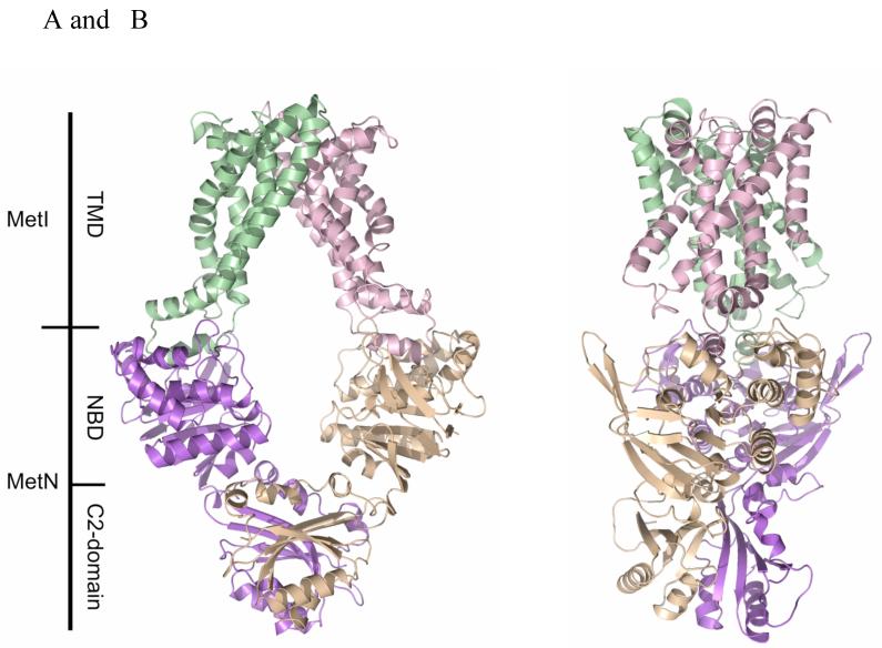
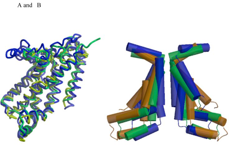
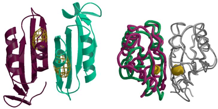
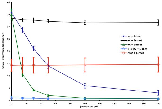
The crystal structure of the high-affinity Escherichia coli MetNI methionine uptake transporter, a member of the adenosine triphosphate (ATP)-binding cassette (ABC) family, has been solved to 3.7 angstrom resolution. The overall architecture of MetNI reveals two copies of the adenosine triphosphatase (ATPase) MetN in complex with two copies of the transmembrane domain MetI, with the transporter adopting an inward-facing conformation exhibiting widely separated nucleotide binding domains. Each MetI subunit is organized around a core of five transmembrane helices that correspond to a subset of the helices observed in the larger membrane-spanning subunits of the molybdate (ModBC) and maltose (MalFGK) ABC transporters. In addition to the conserved nucleotide binding domain of the ABC family, MetN contains a carboxyl-terminal extension with a ferredoxin-like fold previously assigned to a conserved family of regulatory ligand-binding domains. These domains separate the nucleotide binding domains and would interfere with their association required for ATP binding and hydrolysis. Methionine binds to the dimerized carboxyl-terminal domain and is shown to inhibit ATPase activity. These observations are consistent with an allosteric regulatory mechanism operating at the level of transport activity, where increased intracellular levels of the transported ligand stabilize an inward-facing, ATPase-inactive state of MetNI to inhibit further ligand translocation into the cell.


Figures

Figure 1
Figure 1
(A) The ABC transporter MetNI consists of four subunits: two membrane-spanning MetI subunits (green and pink) and two MetN ABC subunits (purple and tan). The molecular rotation axis relating the MetI subunits is vertical, with the cytoplasmic (inward) facing surface of the transporter towards the bottom. The C2-domains forming the dimer interface between MetN subunits are at the bottom. (B) A view of MetNI rotated ∼90° about the vertical axis from that of (A), illustrating the asymmetrical orientation of the C2 domains relative to the MetI subunits and the MetN nucleotide binding domains. This figure was prepared and rendered with PyMOL (38).
Figure 2
Figure 2
(A) Superposition of the five membrane spanning helices forming the common core of the transmembrane domains of MetI (yellow), ModB (green), MalF (blue) and MalG (light blue). TM1 (using the MetI number) is oriented diagonally across the front, while remaining helices are arranged in the sequence TM2 to TM5, from left to right. The coupling helices between TM3 and TM4 are the helical elements at the bottom of the figure. (B) Comparison of the TM2-TM3-TM4 helices lining the translocation pathways of MetI (brown), ModB (green) and MalFG (blue), illustrating the progressive narrowing of cytoplasmic opening of the translocation pathway in the sequence from the methionine to molybdate to maltose transporters. The views in this figure are from the membrane, with the cytoplasmic surface oriented down. Figures 2 and 3 were prepared with MOLSCRIPT and RASTER3D (39, 40).
Figure 3
Figure 3
(A) Anomalous difference Fourier map calculated at 5.2 Å resolution illustrating the binding of selenomethionine to the C2-domains of MetNI following a 1 mM soak of transporter crystals. The electron density is contoured at 6 times the standard deviation of the map. (B) Comparison of the relative orientations of C2-domains observed in the MetNI and free MetN-C2 structures, following superposition of one subunit in each dimer. The green and dark gray traces correspond to the C2-domain dimer in MetNI, while the magenta and light gray traces represent the isolated C2-domain structure. The binding site for selenomethionine is denoted by the gold surface. The view is roughly perpendicular about the horizontal axis from that in (A).
Figure 4
Figure 4
Dependence of the ATPase activity of various MetNI constructs on the concentration of methionine analogues. The ATPase activity was measured from the rate of phosphate production as assayed by the method of Webb (41), and converted to moles phosphate/minute/mole transporter. An initial ATP concentration of 1 mM was used, which is ∼3 times the apparent Km (Figure S3). The blue diamonds, black squares and green triangles correspond to L-methionine, D-methionine and L-selenomethionine, while the open blue and red circles represent the effect of L-methionine on the E166Q (ATPase-inactive mutant) and the ΔC2-MetNI truncation mutant, respectively. The error bars represent the standard deviations calculated from 8 measurements.

References

    1. Kadner RJ. J. Bact. 1974;117:232. - PMC - PubMed
    1. Kadner RJ, Watson WJ. J. Bact. 1974;119:401. - PMC - PubMed
    1. Zhang Z, et al. Arch. Microbiol. 2003;180:88. - PubMed
    1. Higgins CF. Annu. Rev. Cell Biol. 1992;8:67. - PubMed
    1. Holland IB, Cole SPC, Kuchler K, Higgins CF, editors. ABC proteins: From Bacteria to Man. Academic; London: 2003. p. 647.

Publication types

MeSH terms