. 2008 Nov 14;135(4):623-34.
doi: 10.1016/j.cell.2008.09.058.

Structural synergy and molecular crosstalk between bacterial helicase loaders and replication initiators

Affiliations

Structural synergy and molecular crosstalk between bacterial helicase loaders and replication initiators

Melissa L Mott et al. Cell. .

Abstract

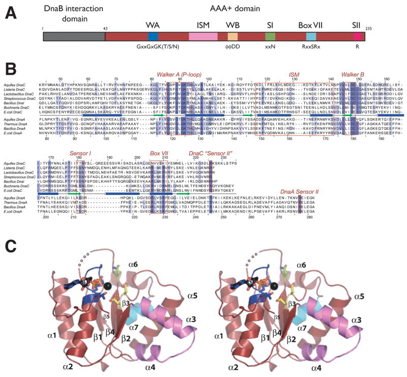
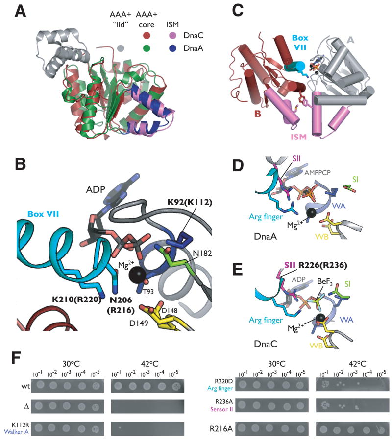
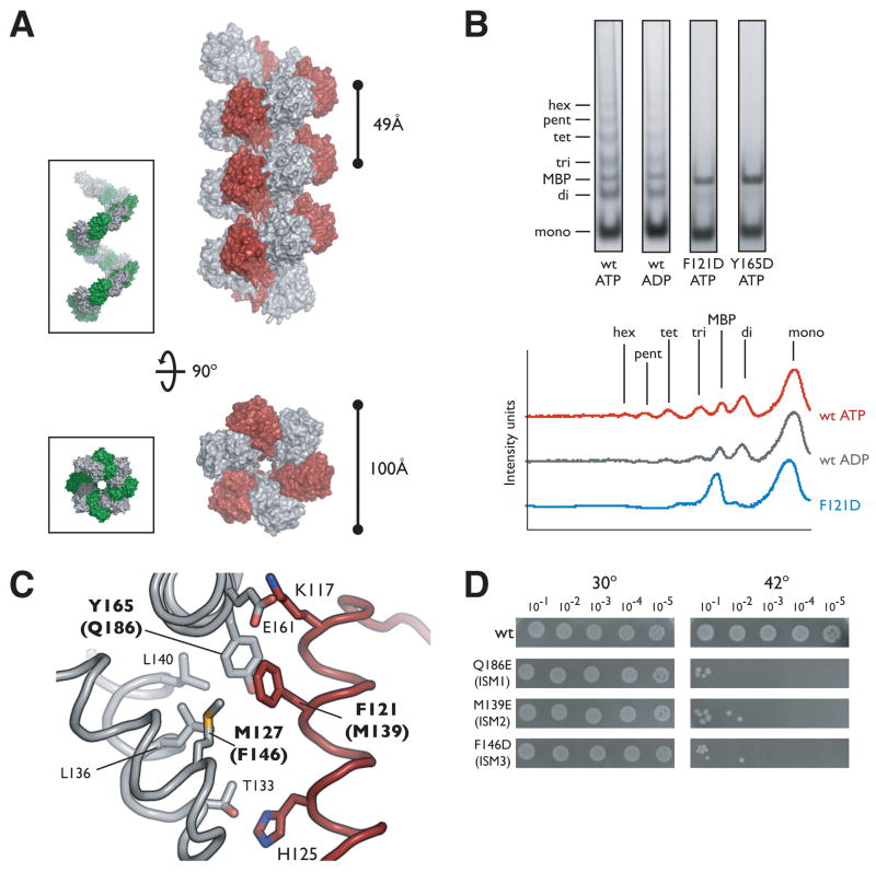
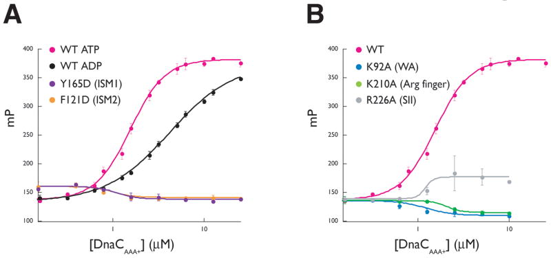
The loading of oligomeric helicases onto replication origins marks an essential step in replisome assembly. In cells, dedicated AAA+ ATPases regulate loading, however, the mechanism by which these factors recruit and deposit helicases has remained unclear. To better understand this process, we determined the structure of the ATPase region of the bacterial helicase loader DnaC from Aquifex aeolicus to 2.7 A resolution. The structure shows that DnaC is a close paralog of the bacterial replication initiator, DnaA, and unexpectedly shares an ability to form a helical assembly similar to that of ATP-bound DnaA. Complementation and ssDNA-binding assays validate the importance of homomeric DnaC interactions, while pull-down experiments show that the DnaC and DnaA AAA+ domains interact in a nucleotide-dependent manner. These findings implicate DnaC as a molecular adaptor that uses ATP-activated DnaA as a docking site for regulating the recruitment and correct spatial deposition of the DnaB helicase onto origins.


Figures

Figure 1
Figure 1. Structure of DnaCAAA+
(A) Domain representation of DnaC. The N-terminal helicase binding region is colored gray and the central AAA+ domain is shown in red. Numbers refer to amino acid positions. AAA+ motifs are highlighted. WA – Walker-A, WB – Walker-B, SI – sensorI, SII – sensor-II, ISM – Initiator Specific Motif. (B) Sequence alignment of selected DnaC and DnaA homologs. Alignment was generated by ClustalX (Thompson et al., 1997). (C) Stereo view of DnaCAAA+. Walker-A and -B motifs are blue and yellow, respectively. The sensor-I residue is green and the Box VII helix cyan. ADP and the coordinating magnesium ion (black) are shown within the ATP binding cleft. An internal disordered region is shown as a dotted line. This and all other molecular figures were generated with PyMOL (pymol.sourceforge.net).
Figure 2
Figure 2. Nucleotide binding by DnaCAAA+
(A) Superposition of DnaCAAA+ and the AAA+ domain from Aquifex aeolicus DnaA (PDB ID 2HCB). (B) Close-up view of the ADP-DnaCAAA+ active site. Residues from two protomers of DnaCAAA+ are shown in gray and red, respectively. The Box VII helix of one molecule is cyan. Residues selected for mutation are labeled in bold; numbering for equivalent residues in E. coli is given in parentheses. (C) Cartoon representation of two associated ADP-DnaCAAA+ protomers, colored gray and red, respectively. The Box VII helix of monomer B is cyan and Lys210 is shown as sticks. The ISMs of both subunits are violet. Residues lining the ISM that were selected for mutational studies are indicated as sticks (see also Figure 3B). (D) The ATP-DnaA active site. AAA+ motifs of the initiator are colored according to the scheme presented for DnaCAAA+ in Figure 1A. (E) The DnaCAAA+ active site in the presence of the ATP mimetic ADP·BeF3. (F) Mutations in DnaC AAA+ motifs cause growth defects in vivo. Cultures of the E. coli dnaC2 strain transformed with a plasmid carrying the indicated DnaC mutant were serially diluted (1:10) and grown overnight on LB/Kan plates at the indicated temperature. wt= wild-type, Δ= empty vector.
Figure 3
Figure 3
DnaCAAA+ oligomerization (A) DnaCAAA+ forms a right-handed filament. Side and axial views of 21 symmetry-related DnaCAAA+ monomers are shown in surface representation with alternating subunits colored gray and red. The inset depicts similar views for the AAA+ domains of the ATP-DnaA filament (PDB ID H2HCB) with alternating monomers in gray and green. (B) DnaCAAA+ forms multimers in solution. Crosslinking was performed by the addition of 1.25mM glutaraldehyde to wild-type (wt) DnaC, or either ISM mutant (F121D and Y165D) in the presence of the indicated nucleotide. The graph on the right compares the intensity traces for wt ATP, wt ADP, and the ISM mutant F121D. mono – monomer, di –dimer, tri – trimer, tet – tetramer, pent – pentamer, hex – hexamer, MBP – maltose binding protein. (C) Close-up view of the interface created by the packing of ISMs from adjacent DnaC AAA+ protomers in the filament. Residues selected for mutation are indicated in bold, the numbering for equivalent residues in E. coli is given in parentheses. (D) Mutation of residues involved in intermolecular DnaCAAA+ contacts result in significant growth defects in vivo. Experiments were performed similarly to those described for Figure 2F.
Figure 4
Figure 4. Single-stranded DNA binding by DnaCAAA+
(A) The effect of nucleotide and interface mutants on DnaC-ssDNA interactions. Binding reactions contained 10nM fluorescein-labeled dT25 oligonucleotide titrated against either wild-type DnaCAAA+ or either interface mutant. All assays were performed in triplicate the presence of 2mM ATP except where indicated. Error bars represent the standard deviation between measurements. (B) AAA+ motif mutations disrupt ssDNA binding by DnaCAAA+. Experiments were performed similarly as for (A) in the presence of 2mM ATP. Data points for wild-type DnaCAAA+ are the same as those indicated in (A), and shown again for comparison.
Figure 5
Figure 5. The AAA+ domains of DnaC and DnaA interact in a nucleotide-dependent manner
(A) Co-precipitation of DnaCAAA+ by domains III and IV of Aquifex aeolicus DnaA fused to maltose binding protein (MBP-DnaAIII–IV). The positions for MBP-DnaAIII–IV, untagged DnaAIII–IV, and DnaCAAA+ on the SDS PAGE gel are indicated. Apo - no nucleotide was added to reaction conditions; “ISM mutant” - double mutant F121D/Y165D. Free MBP present in lane 9 resulted from incomplete separation of the protein from the DnaCAAA+ ISM mutant during purification (see Sup. Material). (B) Quantitation of the results presented in (A). Experiments for each condition were repeated three times and averaged. Error bars represent the variance between experiments.
Figure 6
Figure 6. Model for DnaC/DnaA crosstalk and helicase deposition
(A) Structural model for oligomeric DnaC:DnaA interactions. The figure was generated by superimposing the last subunit of a six-subunit DnaCAAA+ oligomer onto the end of a twelve-subunit DnaA filament assembly. Axial and side views are shown. Cyan spheres represent bound nucleotide. (B) Model for the symmetric loading of two replicative helicases at oriC. Left – DnaA assembles at oriC and melts the DUE (purple strands). Middle, (1) – helicase loading on the bottom DUE strand is facilitated through direct DnaA:DnaB interaction. Middle, (2) - DnaC, through a specific interaction with ATP-charged DnaA, recruits the helicase destined for the top strand to oriC. Right – ATP hydrolysis and loss of DnaC frees both DnaB hexamers to migrate to their proper fork positions.

References

    1. Allen GC, Jr, Kornberg A. Fine balance in the regulation of DnaB helicase by DnaC protein in replication in Escherichia coli. J Biol Chem. 1991;266:22096–22101. - PubMed
    1. Baker TA, Bell SP. Polymerases and the replisome: machines within machines. Cell. 1998;92:295–305. - PubMed
    1. Barcena M, Ruiz T, Donate LE, Brown SE, Dixon NE, Radermacher M, Carazo JM. The DnaB.DnaC complex: a structure based on dimers assembled around an occluded channel. Embo J. 2001;20:1462–1468. - PMC - PubMed
    1. Bell SP, Dutta A. DNA replication in eukaryotic cells. Annu Rev Biochem. 2002;71:333–374. - PubMed
    1. Biswas SB, Flowers S, Biswas-Fiss EE. Quantitative analysis of nucleotide modulation of DNA binding by DnaC protein of Escherichia coli. Biochem J. 2004;379:553–562. - PMC - PubMed

Publication types

MeSH terms

LinkOut - more resources