. 2008 Nov 5;27(21):2918-28.
doi: 10.1038/emboj.2008.208. Epub 2008 Oct 9.

Structural basis of cargo membrane protein discrimination by the human COPII coat machinery

Affiliations

Structural basis of cargo membrane protein discrimination by the human COPII coat machinery

Joseph D Mancias et al. EMBO J. .

Abstract

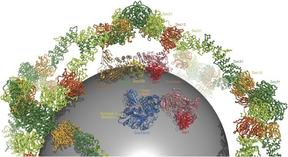
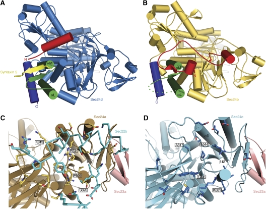
Genomic analysis shows that the increased complexity of trafficking pathways in mammalian cells involves an expansion of the number of SNARE, Rab and COP proteins. Thus, the human genome encodes four forms of Sec24, the cargo selection subunit of the COPII vesicular coat, and this is proposed to increase the range of cargo accommodated by human COPII-coated vesicles. In this study, we combined X-ray crystallographic and biochemical analysis with functional assays of cargo packaging into COPII vesicles to establish molecular mechanisms for cargo discrimination by human Sec24 subunits. A conserved IxM packaging signal binds in a surface groove of Sec24c and Sec24d, but the groove is occluded in the Sec24a and Sec24b subunits. Conversely, LxxLE class transport signals and the DxE signal of VSV glycoprotein are selectively bound by Sec24a and Sec24b subunits. A comparative analysis of crystal structures of the four human Sec24 isoforms establishes the structural determinants for discrimination among these transport signals, and provides a framework to understand how an expansion of coat subunits extends the range of cargo proteins packaged into COPII-coated vesicles.


Figures

Figure 1
Figure 1
Signal sequences on membrin and syntaxin 5 for binding the Sec24c and d isoforms. (A) An isoform-specific packaging assay is shown. Vesicles were generated from semi-intact HEK-293 or CHO-K1 cells using Sar1a, Sec13a/31a and the four isoforms of human Sec24 (Sec23a/Sec24a–d), in the presence and absence of 0.2 mM GTP and an ATP-regenerating system (see Materials and methods). The presence of ERGIC-53, syntaxin 5, ribophorin (HEK-293 cells) and membrin (CHO-K1 cells) in budded vesicles and total membranes (T) was determined by immunoblotting. Polyclonal antibodies to syntaxin 5 recognize both syntaxin 5 splice variants: a 35-kDa form (lower band) and a 42-kDa form (upper band). (B) Localization of the Sec24c-binding site on membrin. SNARE–GST fusion proteins were incubated with an input mixture comprising Sec24c (0.3 mg/ml) and E. coli lysate (13 mg/ml). Specific binding was assessed by the enrichment of Sec24c from the input (lane 2). Bound proteins were analysed by 4–15% SDS–PAGE and Coomassie blue staining. To pinpoint the transport signal sequence on membrin, residues TTIPMD were mutated in turn to alanine in the context of the membrin (107–126)–GST construct (lanes 7–12). (C) Alignment of membrin and syntaxin 5 sequences. Polypeptide sequences of membrin from six organisms (see Materials and methods) were aligned using ClustalW. Just the linker region (that joins the N-terminal domain to the SNARE motif) is shown here; highly conserved residues are outlined in yellow and the key IxM sequence is boxed in red. The alignment of syntaxin 5 from seven organisms (see Materials and methods) also shows just the linker region; highly conserved residues are outlined in yellow, the predicted IxM sequence is boxed in red, and the YNNSNPF and LxxME motifs within S. cerevisiae syntaxin 5 are boxed in blue. (D) Localization of Sec24c-binding site on the syntaxin 5 linker. Residues of the DVAIDMM sequence were mutated to alanine in the context of the syntaxin 5 (181–200)–GST construct (lanes 6–11).
Figure 2
Figure 2
Molecular recognition of syntaxin 5 and membrin IxM packaging signals by Sec24d. (A) Crystal structure of the Sec23a/24d complex viewed from the membrane distal surface. Bound to Sec24d is the transport signal of syntaxin 5 (coloured yellow). (B) This alternate view, approximately along the long axis of the Sec23a/24d complex, emphasizes the proximity of the syntaxin 5 signal to the modelled membrane (grey line). (C) Two views of the syntaxin 5 transport signal bound to Sec24d. The upper picture shows a close-up view of the DVAIDMM peptide of syntaxin 5 bound to the COPII protein; the critical Ile191 and Met193 residues are labelled. The lower picture shows the syntaxin 5 peptide together with a difference electron density map (SA omit map; Brünger, 1998), calculated using the Sec23a/24d·syntaxin 5 data at 2.7-Å resolution and contoured at 2.2σ (Table I). (D) Detailed view of the membrin TTIPMD peptide bound to Sec24d. In the upper picture, the Ile118 and Met120 residues on membrin are labelled. The lower picture shows the membrin transport signal and a difference electron density (SA omit) map, calculated using the Sec23a/24d·membrin data at 3.0-Å resolution and contoured at 2.0σ.
Figure 3
Figure 3
Structural basis for signal-mediated packaging of membrin and syntaxin 5 into COPII vesicles. (A) Molecular surface of the Sec23a/24d complex oriented approximately as in Figure 2B. Sec23a is coloured pink and Sec24d is blue. The IxM-binding site (those residues contacted by the syntaxin 5 transport signal) is highlighted in cyan. The syntaxin 5 peptide is coloured yellow and drawn as an arrow to emphasize its β-strand-type connection to Sec24d. (B) Conservation of the signal-binding site on Sec24d. The molecular surface of Sec24d is coloured according to sequence conservation of the underlying residues in an alignment of Sec24c and Sec24d isoforms from nine organisms (see Materials and methods). The IxM-binding site on Sec24d is outlined in black. (C) Interactions between the IxM signal of membrin and Sec24d. The key residues of membrin, I118 and M120, are labelled. Three central residues of the binding pocket on Sec24d are labelled and boxed: L834, I835 and L836 (the equivalent residues on Sec24c are L895, I896 and L897). (D) In vitro budding assay (performed as in Figure 1A) from semi-intact HEK-293 cells (to monitor packaging of ERGIC-53, syntaxin 5 and ribophorin) and CHO-K1 cells (for membrin). The packaging of cargo was assessed for wild-type Sec24c and the IxM site triple-mutant Sec24c–895AAA897 (This is part of the same experiment as that shown in Figure 1A.). The signal/noise of the blot for syntaxin 5 is limited by the quality of the antibody.
Figure 4
Figure 4
Molecular recognition of Bet1 and VSV-G transport signals at the B site of Sec24a and Sec24b. (A) Interactions were tested between the cytoplasmic tail (29 C-terminal residues) of VSV-G and the four human Sec24 isoforms. VSV-G binds to Sec24a and Sec24b (lanes 4 and 8), but not to Sec24c or Sec24d (lanes 12 and 16). Mutation of the aspartate and glutamate residues of the VSV-G DxE motif abrogates binding to Sec24a (lanes 19 and 21); the intervening isoleucine residue can be mutated to alanine without appreciable effect on binding (lane 20). These pull-down experiments were performed as in Figure 1B. (B) Crystal structure of the Sec23a/24a·Sec22b complex, with the membrane-proximal surface facing forward. Circled in the picture is the VSV-G transport signal bound to the B site on Sec24a. Note the proximity of the VSV-G- and Sec22b-binding sites on Sec24a. (C) Interactions between the DxE signal on VSV-G and the B site of Sec24a. Difference electron density for the bound VSV-G peptide (QIYTDIEMNR) was calculated at 2.7-Å resolution and contoured at 1.8σ. (D) Interactions between the Bet1 signal and the B site of Sec24a. Difference electron density for the bound Bet1 peptide (GYSACEEEN) was calculated at 3.3-Å resolution and contoured at 1.7σ.
Figure 5
Figure 5
Arrangement of cargo-binding sites in the COPII coat. In the figure, the model of the Sec13/31 cage is drawn as a backbone worm and coloured red and green following the convention of Fath et al (2007). Modelled inside the cage is a sphere of diameter 40 nm representing a membrane vesicle. The two copies of the Sec23/24·Sar1 pre-budding complex are composite models based on structural alignments of crystal structures from the current study and from previous studies (Bi et al, 2002; Mossessova et al, 2003). Sec23a is coloured pink, Sar1 (taken from the yeast Sec23/24·Sar1 complex) is red, Sec24a is brown and Sec24d is blue. The bound cargo molecules are coloured as follows: VSV-G/Bet1 is green, Sec22b is yellow, the binding site for yeast Sed5 is labelled with a star in orange and syntaxin 5/membrin is yellow.
Figure 6
Figure 6
Structural basis for cargo discrimination by human Sec24 isoforms. (A) Crystal structure of the Sec23a/Sec24d·syntaxin 5 complex viewed from the base of Sec24d. The αL–αM loop that forms the binding site for IxM signals is coloured green, the C-terminal helix is deep blue and the variable N-terminal region is red. The syntaxin 5 peptide is yellow. (B) Crystal structure of Sec24b (see Table I). As in (A), the αL–αM loop is coloured green, the C-terminal helix is deep blue and the N-terminal region is red. Note how the N-terminal variable region occupies the IxM-binding site. (C) Structure of Sec24a from the Sec23a/Sec24a·Sec22b complex. Sec24a is coloured brown and Sec23a is pink. Sec24a residues that form the Sec22-binding site are drawn in yellow. Critical interface residues R541 and P500 are labelled. (D) Crystal structure of Sec24c. Sec24c is coloured light blue and Sec23a is modelled in pink (Sec23a is not present in this crystal structure; Table I). Sec24c cannot bind to Sec22 because key binding-site residues are altered: in particular, note that residue 544 is alanine (equivalent to R541 in Sec24a) and residue 498 is phenylalanine (P500 in Sec24a).

References

    1. Bi X, Corpina RA, Goldberg J (2002) Structure of the Sec23/24–Sar1 pre-budding complex of the COPII vesicle coat. Nature 419: 271–277 - PubMed
    1. Bi X, Mancias JD, Goldberg J (2007) Insights into COPII coat nucleation from the structure of Sec23·Sar1 complexed with the active fragment of Sec31. Dev Cell 13: 635–645 - PMC - PubMed
    1. Bock JB, Matern HT, Peden AA, Scheller RH (2001) A genomic perspective on membrane compartment organization. Nature 409: 839–841 - PubMed
    1. Bonifacino JS, Glick BS (2004) The mechanisms of vesicle budding and fusion. Cell 116: 153–166 - PubMed
    1. Bracher A, Weissenhorn W (2002) Structural basis for the Golgi membrane recruitment of Sly1p by Sed5p. EMBO J 21: 6114–6124 - PMC - PubMed

Publication types

MeSH terms