. 2009 Jun 12;324(5933):1444-7.
doi: 10.1126/science.1170481.

Structure of rotavirus outer-layer protein VP7 bound with a neutralizing Fab

Affiliations

Structure of rotavirus outer-layer protein VP7 bound with a neutralizing Fab

Scott T Aoki et al. Science. .

Abstract

Rotavirus outer-layer protein VP7 is a principal target of protective antibodies. Removal of free calcium ions (Ca2+) dissociates VP7 trimers into monomers, releasing VP7 from the virion, and initiates penetration-inducing conformational changes in the other outer-layer protein, VP4. We report the crystal structure at 3.4 angstrom resolution of VP7 bound with the Fab fragment of a neutralizing monoclonal antibody. The Fab binds across the outer surface of the intersubunit contact, which contains two Ca2+ sites. Mutations that escape neutralization by other antibodies suggest that the same region bears the epitopes of most neutralizing antibodies. The monovalent Fab is sufficient to neutralize infectivity. We propose that neutralizing antibodies against VP7 act by stabilizing the trimer, thereby inhibiting the uncoating trigger for VP4 rearrangement. A disulfide-linked trimer is a potential subunit immunogen.


Figures
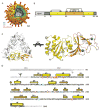
Fig. 1
Fig. 1
Structure of rhesus rotavirus VP7. A. Structure of the complete virion as determined by cryoEM and filtered at 25 Å resolution. The segmentation of the structure is based on reconstructions of the complete virion (Settembre et al, in preparation) and the VP7-coated DLP (Chen et al, submitted) and on published work of others (refs). B. Schematic diagram of the VP7 primary structure, including the signal sequence (residues 1–50, light gray). The two domains are in the same colors as in A; the N- and C-terminal arms, disordered in the crystal structure, are in dark gray. The pattern of intrasubunit disulfide bonds is also shown, with numbers corresponding to the positions of the cysteine residues. C. Ribbon diagram of the trimer (left), viewed along its threefold axis (i.e., as if looking onto the surface of the virion) with one subunit and one pair of Ca2+ ions in color and the other two subunits in gray. The Rossmann-fold domain (domain I) is in yellow; the β-barrel domain (domain II), in orange; the Ca2+ ions, in blue. A single subunit, in side view, is shown on the right, with secondary structure elements labeled. D. Amino-acid sequence (single-letter code) of rhesus rotavirus VP7. Helices and strands are shown as cylinders and arrows, above the corresponding sequence. Colored, underlined residue letters are the positions of neutralization escape mutations, as described in Fig. 4 and Table S2. (See Fig. 4 caption for color code.) Dark blue letters, not underlined, are residues that contribute to Ca2+ ion ligation (either through side-chain groups or through main-chain carbonyls): see Fig. 2. Scissors symbol: position of signal peptidase cleavage. Branched symbol at Asn69: position of N-linked glycan.
Fig. 2
Fig. 2
View of the subunit interface, showing Ca2+ ion binding sites (labeled as 1 and 2) and the Fab contact. The view is from a direction that would be 60° around to the right in the right-hand panel of Fig. 1C. Residues that contribute to Ca2+ ligation (through side-chain groups or main-chain carbonyls) are included in stick representation and labeled. The light chain is in red; the heavy chain, in cyan. VP7 colors, as in Fig. 1. The light-chain CDR3, the heavy-chain CDR2, and several secondary-structure elements in VP7 are labeled, to provide points of reference and to facilitate comparison with Fig. 1. The heavy-chain CDR3, which like the light-chain CDR3 has extensive contacts with VP7, is the loop in the rear with a projected figure-eight-like appearance.
Fig. 3
Fig. 3
Neutralization of RRV by the IgG and Fab of mAb 4F8. Percent infectivity is plotted (on a log scale) against antibody (or Fab) concentration (also on a log scale). Error bars calculated are from three independent measurements. For details of the assay, see supporting on-line materials.
Fig. 4
Fig. 4
Positions of neutralization escape mutations selected by various monoclonal antibodies. The residues cluster roughly into two regions, designated 7-1 and 7-2 (see text). Region 7-1 spans the intersubunit boundary; we have divided it into 7-1a (red), on one side of the interface, and 7-1b (pink), on the other. Residues in 7-2 are in blue. The 4F8 Fab (and by inference the Fabs of most mAbs the recognize residues in region 7-1) has contacts in both 7-1a and 7-1b (see Fig. 2). Positions at which escape is through a mutation that produces a new glycosylation site are in orange and lie either in 7-1 or 7-2; an asterisk labels residue 211, which is mentioned in the text.

References

    1. Estes MK, Kapikian AZ. In: Fields Virology. 5. Howley DMKPM, editor. Lippincott, Williams & Wilkins; Philadelphia: 2007. pp. 1918–1974.
    1. Svensson L, Dormitzer PR, von Bonsdorff CH, Maunula L, Greenberg HB. J Virol. 1994;68:5204. - PMC - PubMed
    1. Lopez T, et al. J Virol. 2005;79:184. - PMC - PubMed
    1. Michelangeli F, Liprandi F, Chemello ME, Ciarlet M, Ruiz MC. J Virol. 1995;69:3838. - PMC - PubMed
    1. Dormitzer PR, Greenberg HB, Harrison SC. Virology. 2000;277:420. - PubMed

Publication types

MeSH terms

Associated data