. 2010 Jul 8;466(7303):272-5.
doi: 10.1038/nature09136.

Structural basis for the coupling between activation and inactivation gates in K(+) channels

Affiliations

Structural basis for the coupling between activation and inactivation gates in K(+) channels

Luis G Cuello et al. Nature. .

Abstract

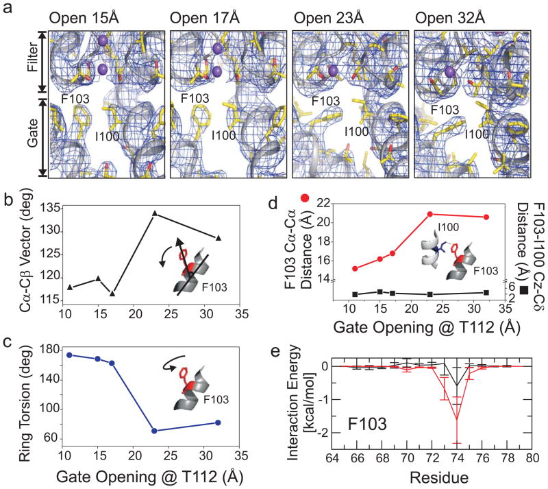
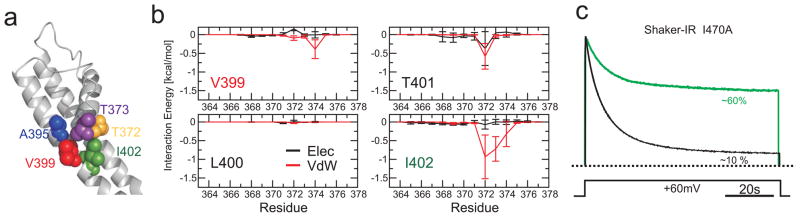
The coupled interplay between activation and inactivation gating is a functional hallmark of K(+) channels. This coupling has been experimentally demonstrated through ion interaction effects and cysteine accessibility, and is associated with a well defined boundary of energetically coupled residues. The structure of the K(+) channel KcsA in its fully open conformation, in addition to four other partial channel openings, richly illustrates the structural basis of activation-inactivation gating. Here, we identify the mechanistic principles by which movements on the inner bundle gate trigger conformational changes at the selectivity filter, leading to the non-conductive C-type inactivated state. Analysis of a series of KcsA open structures suggests that, as a consequence of the hinge-bending and rotation of the TM2 helix, the aromatic ring of Phe 103 tilts towards residues Thr 74 and Thr 75 in the pore-helix and towards Ile 100 in the neighbouring subunit. This allows the network of hydrogen bonds among residues Trp 67, Glu 71 and Asp 80 to destabilize the selectivity filter, allowing entry to its non-conductive conformation. Mutations at position 103 have a size-dependent effect on gating kinetics: small side-chain substitutions F103A and F103C severely impair inactivation kinetics, whereas larger side chains such as F103W have more subtle effects. This suggests that the allosteric coupling between the inner helical bundle and the selectivity filter might rely on straightforward mechanical deformation propagated through a network of steric contacts. Average interactions calculated from molecular dynamics simulations show favourable open-state interaction-energies between Phe 103 and the surrounding residues. We probed similar interactions in the Shaker K(+) channel where inactivation was impaired in the mutant I470A. We propose that side-chain rearrangements at position 103 mechanically couple activation and inactivation in KcsA and a variety of other K(+) channels.


Figures

Figure 1
Figure 1
Conformational coupling between activation and inactivation gates in K+ channels. a. Defining coupling from the selectivity filter to the activation gate by modulating the rate and extent of inactivation. Left panel, KcsA macroscopic currents obtained at 100 mV from liposome-reconstituted KcsA in 100 mM symmetric KCl (red), 100 mM symmetric RbCl (navy blue) or the mutant E71A in 100 mM symmetric KCl (light blue). The right panel shows the extent of inner gate opening for these three conditions in KcsA spin labeled at G116C, and monitored from the amplitude of the central resonance line normalized by the total number of spins. Asolectin-reconstituted spin-labeled channels in 100 mM symmetric KCl (red circles), 100 mM symmetric RbCl (navy blue circles) or the mutant E71A in 100 mM symmetric KCl (light blue circles). b. Defining coupling from the activation gate to the selectivity filter by modulating the extent of inner gate opening. Left, macroscopic currents at 100 mV and 100 mM symmetric KCl for wild-type KcsA (red) or Chymotrypsin C-terminal truncated channel KcsA Δ-125. Right panel, EPR spectra from the spin labeled Y82C mutant in the closed, conductive state (pH 7, black trace); open, inactivated state (pH 3, blue trace); and C-terminal truncated open, inactivated state (pH 3, red trace). EPR spectra were normalized by the total number of spins in the sample.
Figure 2
Figure 2
Structural basis for allosteric coupling in KcsA. a. Conformational rearrangements in the KcsA aqueous cavity in a series of partially opened structures. In each panel, the composite omit map (contoured at 2σ) of the channel corresponding to two neighbouring subunits is shown as a blue mesh. Transmembrane helices are represented as ribbons, and residues Ile100 and Phe103 are shown in stick representation, while ions are shown as space filling spheres. Dependence of the Phe103 side chain tilting (b.), Torsional angle (c.) Cα-Cα inter-subunit distance, and distance between F103 and Ile100 from adjacent subunits (d.) with the degree of opening of the inner bundle gate at Thr112. e. Interaction energies of residue F103 in TM2 with individual side chains from residues in the pore helix.
Figure 3
Figure 3
Role of Phe103 in allosteric coupling with the selectivity filter. a. The mutation F103A sharply reduces the rate and extent of C-type inactivation. Left, representative single channel recordings from asolectin reconstituted channels in symmetric 100 mM KCl. Right, the extent of steady-state inactivation from the ratio between peak (Ipeak) and steady state (I) current plotted as a function of side chain volume for four substitutions at position 103. Multiple side-chain substitutions at position 103 show a volume dependence of the extent of inactivation, as derived from macroscopic currents during pH pulse experiments (inset). b. Mutation of Phe103 disrupts C-type inactivation in the deeply inactivated mutant E71H. Top, representative single channel recordings in symmetric 100 mM KCl from the E71H mutant background and the E71H-F103A double mutant. Bottom, crystal structure of E71H-F103A KcsA, determined at 3 Å resolution. Shown are the 2Fo−Fc electron density map (magenta mesh contoured at 2σ) of the filter corresponding to residues 70–80 from two diagonally symmetric subunits and the Fo−Fc omit map (cyan mesh contoured at 3σ) corresponding to ions in the filter.
Figure 4
Figure 4
A common gate coupling mechanism in K+ channels. a. Structural representation of the interface between the inner bundle gate (S6) and the pore helix in the Kv 1.2 pore domain (2R9R) with key amino acid side chains shown as van der Waals spheres (Equivalent KcsA/Kv 1.2 positions: Met 96/Ala391, blue; Ile100/Val399, Red; and Phe103/Ile401, green; T74/T373, yellow; T75/T374, purple). b. Interaction free energies of selected Kv1.2 residues in TM2 (equivalent to those in KcsA) with individual side chains from residues in the pore helix. c. Equivalent role of Ile470 in gate coupling in Shaker. Representative Cut-open oocyte voltage clamp traces obtained from N-type inactivation-removed Shaker (Shaker-IR), black trace and the mutation I470A (green trace) expressed in Xenopus oocytes (n=5 for I470A). Currents were normalized to the peak current.

References

    1. Panyi G, Deutsch C. Cross talk between activation and slow inactivation gates of Shaker potassium channels. J Gen Physiol. 2006;128:547–559. - PMC - PubMed
    1. Sadovsky E, Yifrach O. Principles underlying energetic coupling along an allosteric communication trajectory of a voltage-activated K+ channel. Proc Natl Acad Sci U S A. 2007;104:19813–19818. - PMC - PubMed
    1. Baukrowitz T, Yellen G. Use-dependent blockers and exit rate of the last ion from the multi-ion pore of a K+ channel. Science. 1996;271:653–656. - PubMed
    1. Lopez-Barneo J, Hoshi T, Heinemann SH, Aldrich RW. Effects of external cations and mutations in the pore region on C-type inactivation of Shaker potassium channels. Receptors Channels. 1993;1:61–71. - PubMed
    1. Cuello L, Jogini V, Perozo E. Structural mechanism of C-type inactivation in K+ channnels. Nature. 2010 In Press. - PMC - PubMed

Publication types

MeSH terms