Review
. 2018 Jul 6;293(27):10512-10523.
doi: 10.1074/jbc.TM117.000374. Epub 2017 Dec 14.

Nonhomologous DNA end-joining for repair of DNA double-strand breaks

Affiliations
Review

Nonhomologous DNA end-joining for repair of DNA double-strand breaks

Nicholas R Pannunzio et al. J Biol Chem. .

Abstract

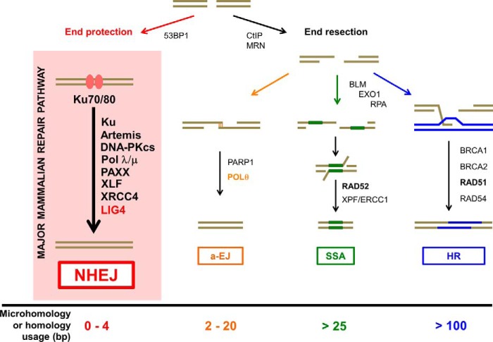
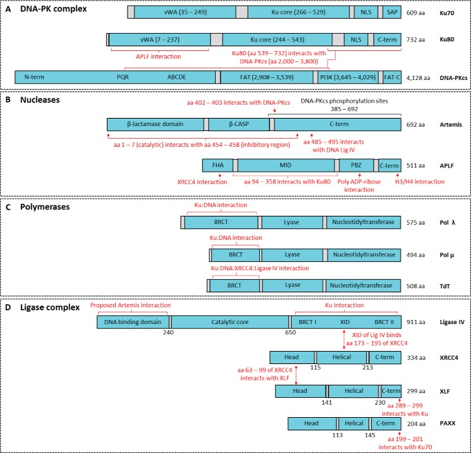
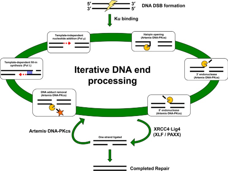
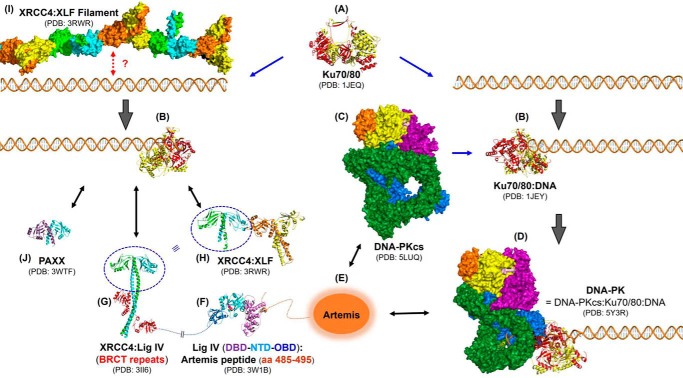
Nonhomologous DNA end-joining (NHEJ) is the predominant double-strand break (DSB) repair pathway throughout the cell cycle and accounts for nearly all DSB repair outside of the S and G2 phases. NHEJ relies on Ku to thread onto DNA termini and thereby improve the affinity of the NHEJ enzymatic components consisting of polymerases (Pol μ and Pol λ), a nuclease (the Artemis·DNA-PKcs complex), and a ligase (XLF·XRCC4·Lig4 complex). Each of the enzymatic components is distinctive for its versatility in acting on diverse incompatible DNA end configurations coupled with a flexibility in loading order, resulting in many possible junctional outcomes from one DSB. DNA ends can either be directly ligated or, if the ends are incompatible, processed until a ligatable configuration is achieved that is often stabilized by up to 4 bp of terminal microhomology. Processing of DNA ends results in nucleotide loss or addition, explaining why DSBs repaired by NHEJ are rarely restored to their original DNA sequence. Thus, NHEJ is a single pathway with multiple enzymes at its disposal to repair DSBs, resulting in a diversity of repair outcomes.

Keywords: DNA endonuclease; DNA repair; DNA-dependent serine/threonine protein kinase (DNA-PK); NHEJ; double-stranded DNA breaks; nucleic acid enzymology; protein structure.


Conflict of interest statement

The authors declare that they have no conflicts of interest with the contents of this article

Figures

Figure 1.
Figure 1.
NHEJ in the context of other double-strand break repair pathways. DNA double-strand breaks (DSBs) can be repaired by NHEJ, alternative end-joining (a-EJ), single-strand annealing (SSA), or homologous recombination (HR). Pathway choice and pathways other than NHEJ are discussed in other Minireviews in this thematic series. The name NHEJ originally arose to distinguish it from repair that requires extensive DNA homology (i.e. HR and SSA). Lengths of terminal microhomology (MH) between 1 and 4 bp are common in NHEJ. a-EJ is also called microhomology-mediated end joining (MMEJ) or Pol θ-mediated end joining (TMEJ). The major difference in the pathways is the requirement for significant DNA end resection. The p53-binding protein 1 (53BP1) is a chromatin remodeler and a positive regulator for NHEJ. Although Artemis·DNA-PKcs can carry out some nucleolytic resection (typically <20 nt), the NHEJ pathway does not require extensive end resection, and the ends are protected from deeper resection by the binding of the Ku heterodimer (Ku70–80) to the DNA ends. By contrast, the C-terminal binding protein-interacting protein (CtIP) and the MRN (MRE11 (meiotic recombination 11)·RAD50·NBS1 (Nijmegen breakage syndrome protein 1)) complexes are involved in extensive 5′ to 3′ resection of regions of the duplex, and this generates stretches of ssDNA at DNA ends for a-EJ, SSA, and HR. SSA typically requires >25 bp of microhomology, whereas the requirement for a-EJ is typically <20 bp. Poly(ADP-ribose) polymerase 1 (PARP1) and Pol θ are important for a-EJ. Bloom syndrome RecQ-like helicase (BLM) and exonuclease 1 (EXO1) account for additional resection, and replication protein A (RPA) binds to ssDNA to promote the SSA and HR pathways. RAD52-mediated annealing of homologous sequence is key for the SSA pathway. XPF-ERCC1 cuts the remaining 3′ nonhomologous ssDNA prior to ligation by DNA ligase 1. By contrast, RAD51-mediated strand exchange with its association with BRCA1, BRCA2, and RAD54 is essential for facilitating the HR pathway.
Figure 2.
Figure 2.
Nonhomologous end-joining proteins and their known interactions. A, nonhomologous end-joining (NHEJ) DNA-dependent protein kinase (DNA-PK) complex consists of a heterodimer of Ku70 and Ku80 plus DNA-PKcs (catalytic subunit). Ku70 and Ku80 consist of von Willebrand (vWA) domains, the Ku core, and the nuclear localization sequence (NLS). Ku70 also contains a SAF-A/B, Acinus, and PIAS (SAP) domain. DNA-PKcs consists of an N-terminal domain with PQR and ABCDE autophosphorylation clusters implicated in its activation, FAT (FRAP, ATM, TRRAP) domain, followed by the phosphatidylinositol 3-kinase (PI3K) domain, and the FAT-C (C-terminal) domain. B, NHEJ nucleases consist of Artemis and APLF (abbreviation for Aprataxin and PNKP-like factor). Artemis has a catalytic β-lactamase domain, a cleavage and polyadenylation specificity factor (β-CASP) domain, and a disordered C terminus. Amino acids (aa) 454–458 bind aa 1–7 to auto-inhibit Artemis activity (119). APLF consists of a forkhead-associated (FHA) domain, middle (MID) domain, and the poly(ADP-ribose)-binding zinc finger (PBZ) domain (73, 120–122). C, polymerases involved in NHEJ are Pol λ, Pol μ, and terminal deoxynucleotidyltransferase (TdT). They consist of a breast cancer C terminus (BRCT) domain, a lyase domain, and a nucleotidyltransferase domain. D, DNA ligase complex consists of DNA ligase IV, X-ray repair cross-complementing 4 (XRCC4), XRCC4-like factor (XLF), and paralog of XRCC4 and XLF (PAXX). DNA ligase IV consists of an N-terminal DNA-binding domain, a catalytic core, and an XRCC4 interaction domain (XID) flanked by the BRCT I and II domains. XRCC4, XLF, and PAXX are structurally similar with an N-terminal head domain, helical domain, and C terminus. Protein domains are in blue and linker regions in gray.
Figure 3.
Figure 3.
DNA ends undergo iterative processing during NHEJ. NHEJ is single pathway with multiple components available to process the diversity of DNA end configurations at any given DSB. The first major step following formation of either a pathological or physiological DSB is binding of the Ku70·Ku80 complex (Ku) to protect DNA ends. The Ku·DNA complex is able to efficiently bind and thereby recruit other NHEJ components. An iterative processing occurs to make the two broken DNA ends optimal for ligation. Several types of processing performed by the Artemis·DNA-PKcs complex or DNA polymerases are shown in the white boxes along the large green circle. It would be difficult to represent all the possible DNA end configurations and every type of enzymatic processing in one figure; therefore, this depiction is not meant to be comprehensive but is merely to highlight some of the possibilities with the key components for each process indicated in parentheses. Any of these processes can occur to either end of a break in any order and multiple times. Once XRCC4·Lig4 is able to successfully ligate across a break, an intermediate with one strand ligated can form. Ligation of the second strand will complete repair. Alternatively, the gapped intermediate generated by ligating one strand has two ss–dsDNA boundaries, and Artemis·DNA-PKcs can cut at either boundary to generate a new DSB, thereby returning the ends to the iterative processing step where they can undergo further alterations.
Figure 4.
Figure 4.
Structural aspects of NHEJ. Select reported three-dimensional structures of NHEJ components are shown. Protein structures are shown in ribbon representation except for DNA-PKcs and an XRCC4·XLF filament, which are shown in surface representations. A, crystal structure of Ku70/80 heterodimer alone at 2.7 Å (PDB code 1JEQ) is shown in the middle (109). Ku70 and Ku80 are shown in red and yellow, respectively. B, these toroidal Ku70/80 proteins bind to broken dsDNA ends to form a Ku70/80·DNA complex (solved at 2.5 Å) (PDB code 1JEY) (109). C, crystal structure of DNA-PKcs at 4.3 Å is shown in the center (PDB code 5LUQ) (84). The DNA-PKcs is color-coded as follows: N terminus (blue); circular cradle (green); head comprising FAT region (purple); kinase (yellow); FRB (orange); FATC (light pink). D, DNA-PKcs binds the Ku70/80·DNA to form a DNA-PK complex. A 6.6-Å cryo-EM structure of DNA-PK holoenzyme is shown (PDB code 5Y3R) (111). E, structure of Artemis has not been reported yet. F, crystal structure of the catalytic region of DNA ligase IV (DBD-NTD-OBD) in complex with an Artemis fragment (aa 485–495) was solved at 2.4 Å (PDB code 3W1B) (94). The Artemis fragment is shown in orange and interacts with the DNA-binding domain (DBD), which is shown in violet. The nucleotidyltransferase domain (NTD) is shown in cyan. The catalytic lysine (Lys-273), which forms a covalent AMP-lysine intermediate, is shown as a sphere, and a possible αPO4 is attached to the lysine. The OB-fold domain (OBD) is shown in blue. G, crystal structure of the complex of XRCC4 homodimer and the BRCT repeats of ligase IV (Lig IV) at 2.4 Å is shown (PDB code 3II6) (107). Each XRCC4 molecule is shown in cyan and green. Two BRCT domains are shown in red. H, crystal structure of XRCC4(1–224)·XLF(1–157) complex (both are homodimers) at 3.94 Å is shown (PDB code 3RWR) (110). The XRCC4 homodimer is shown in cyan and green. The XLF homodimer is shown in yellow and orange. I, this XRCC4·XLF complex can form filaments, shown in the same color scheme at the left top corner, which might bridge DNA ends. J, crystal structure of PAXX homodimer at 3.45 Å is shown in cyan and purple (PDB code 3WTF) (64). Note that Ku70/80 bound on the DNA end can recruit XRCC4·ligase IV complex, and Ku70/80 also directly interacts with and recruits XLF and PAXX through their C termini. Also note that structures of the Pol X family polymerases are not shown here due to a space limitation. The figure was created using PyMOL (The PyMOL Molecular Graphics System, Version 2.0 Schrödinger, LLC).

References

    1. Lieber M. R. (2016) Mechanisms of human lymphoid chromosomal translocations. Nat. Rev. Cancer 16, 387–398 10.1038/nrc.2016.40 - DOI - PMC - PubMed
    1. Moore J. K., and Haber J. E. (1996) Cell cycle and genetic requirements of two pathways of nonhomologous end-joining repair of double-strand breaks in S. cerevisiae. Mol. Cell. Biol. 16, 2164–2173 10.1128/MCB.16.5.2164 - DOI - PMC - PubMed
    1. Meek K., Dang V., and Lees-Miller S. P. (2008) DNA-PK: the means to justify the ends? Adv. Immunol. 99, 33–58 10.1016/S0065-2776(08)00602-0 - DOI - PubMed
    1. Ma Y., Pannicke U., Schwarz K., and Lieber M. R. (2002) Hairpin opening and overhang processing by an Artemis:DNA-PKcs complex in V(D)J recombination and in nonhomologous end joining. Cell 108, 781–794 10.1016/S0092-8674(02)00671-2 - DOI - PubMed
    1. Chang H. H., Watanabe G., Gerodimos C. A., Ochi T., Blundell T. L., Jackson S. P., and Lieber M. R. (2016) Different DNA end configurations dictate which NHEJ components are most important for joining efficiency. J. Biol. Chem. 291, 24377–24389 10.1074/jbc.M116.752329 - DOI - PMC - PubMed

Publication types