. 2012 Nov 6;109(45):18431-6.
doi: 10.1073/pnas.1211702109. Epub 2012 Oct 22.

Structure of Sputnik, a virophage, at 3.5-Å resolution

Affiliations

Structure of Sputnik, a virophage, at 3.5-Å resolution

Xinzheng Zhang et al. Proc Natl Acad Sci U S A. .

Abstract

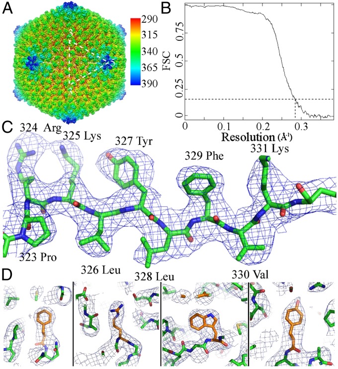
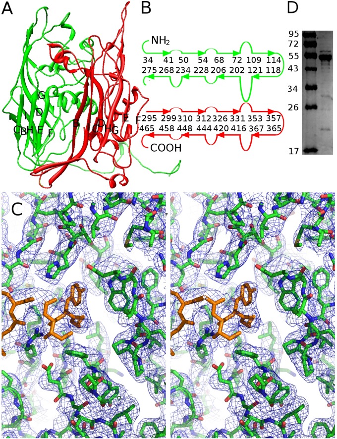
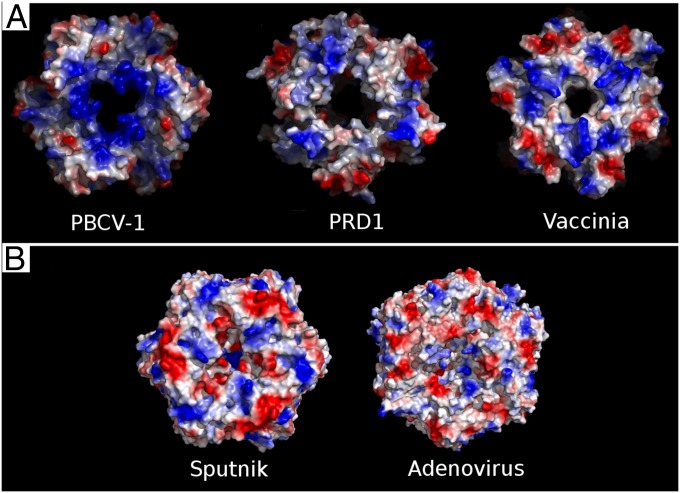
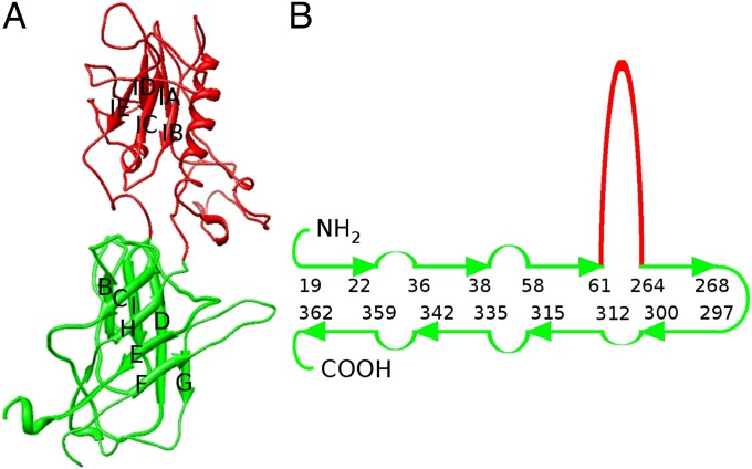
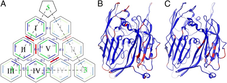
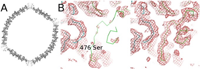
"Sputnik" is a dsDNA virus, referred to as a virophage, that is coassembled with Mimivirus in the host amoeba. We have used cryo-EM to produce an electron density map of the icosahedral Sputnik virus at 3.5-Å resolution, sufficient to verify the identity of most amino acids in the capsid proteins and to establish the identity of the pentameric protein forming the fivefold vertices. It was also shown that the virus lacks an internal membrane. The capsid is organized into a T = 27 lattice in which there are 260 trimeric capsomers and 12 pentameric capsomers. The trimeric capsomers consist of three double "jelly-roll" major capsid proteins creating pseudohexameric capsomer symmetry. The pentameric capsomers consist of five single jelly-roll proteins. The release of the genome by displacing one or more of the pentameric capsomers may be the result of a low-pH environment. These results suggest a mechanism of Sputnik DNA ejection that probably also occurs in other big icosahedral double jelly-roll viruses such as Adenovirus. In this study, the near-atomic resolution structure of a virus has been established where crystallization for X-ray crystallography was not feasible.


Conflict of interest statement

The authors declare no conflict of interest.

Figures

Fig. 1.
Fig. 1.
Reconstruction of Sputnik at 3.5-Å resolution. (A) Reconstruction of the whole virus, colored according to radius. (B) Fourier shell correlation (FSC) coefficient plot showing the resolution of Sputnik reconstruction to be 3.5 Å (FSC = 0.143) according to the criterion defined by Rosenthal and Henderson (42). (C) Cryo-EM density of a β-strand in the MCP. (D) Cryo-EM density of selected residues, Phe, Arg, Trp, and Tyr (from left to right), showing the quality of the reconstruction.
Fig. 2.
Fig. 2.
Double jelly-roll structure of MCP. (A) Ribbon diagram of the Sputnik MCP. The first and second jelly-roll domains are colored green and red, respectively. (B) Diagrammatic representation of the arrangement of β-strands and residue numbers at the ends of the β-strands. (C) Stereo diagram showing the C-terminal region of the MCP. Residues 502–508 are colored orange. These residues bind into a hydrophobic pocket. There is no density or space for the remaining 87 C-terminal residues of the MCP sequence, suggesting that these residues had been cleaved. (D) Molecular mass of the MCP is estimated to be approximately 55 kDa by SDS/PAGE gel.
Fig. 3.
Fig. 3.
Comparison of the inner surface charge distribution of dsDNA virus MCPs with a double jelly-roll fold. (A) Viruses that have a lipid membrane envelope. (B) Viruses that do not have a lipid membrane envelope.
Fig. 4.
Fig. 4.
Single jelly-roll structure of sputnik penton protein. (A) Ribbon diagram of the Sputnik penton protein. The jelly-roll domain is colored green, and the insertion domain is colored red. (B) Diagrammatic representation of the arrangement of β-strands in the penton protein and the residue numbers at the ends of the β-strands.
Fig. 5.
Fig. 5.
Asymmetric unit of Sputnik. (A) Capsomers are identified by Roman numerals I to V. Thirteen independent monomers are identified by Arabic numerals 1 to 13. Each MCP is shown by a green line (the first jelly-roll domain) followed by a blue line (the second jelly-roll domain). The independent minor proteins in the asymmetric unit are colored red. Icosahedrally related positions are indicated in the same manner but with corresponding faded colors. (B) The backbone of the MCP was colored according to the root mean square deviation of each Cα position from the mean of the superimposed 13 independent MCPs, ranging from blue (<0.6 Å) to red (>0.6 Å). (C) Number of times a given residue in the 13 independent MCPs is involved in capsomer to capsomer contacts ranging from blue (<8) to red (>8).
Fig. 6.
Fig. 6.
Structure of the empty Sputnik. (A) Cross-section through the empty Sputnik cryo-EM map, showing where the density is at a higher level. The density of the penton protein is lower than that of the MCPs. (B) Electron density (red) of the hexon capsomer, adjacent to the penton protein in the empty virus (Left) and full virus (Right). The Cα backbone structure (green) of MCP #1, closest to the penton. The electron density from residue 476 to residue 508 of MCP #1 is missing in the empty virus. The Cα backbone structure of penton protein is colored blue.

References

    1. Raoult D, et al. The 1.2-megabase genome sequence of Mimivirus. Science. 2004;306(5700):1344–1350. - PubMed
    1. La Scola B, et al. The virophage as a unique parasite of the giant mimivirus. Nature. 2008;455(7209):100–104. - PubMed
    1. Parola P, et al. Acanthamoeba polyphaga mimivirus virophage seroconversion in travelers returning from Laos. Emerg Infect Dis. 2012;18(9):1500–1502. - PMC - PubMed
    1. Krupovic M, Cvirkaite-Krupovic V. Virophages or satellite viruses? Nat Rev Microbiol. 2011;9(11):762–763. - PubMed
    1. Desnues C, Raoult D. Inside the lifestyle of the virophage. Intervirology. 2010;53(5):293–303. - PubMed

Publication types

Substances

Associated data