. 2010 Apr;17(4):497-503.
doi: 10.1038/nsmb.1792. Epub 2010 Mar 28.

Structural characterization of a capping protein interaction motif defines a family of actin filament regulators

Affiliations

Structural characterization of a capping protein interaction motif defines a family of actin filament regulators

Maria Hernandez-Valladares et al. Nat Struct Mol Biol. 2010 Apr.

Abstract

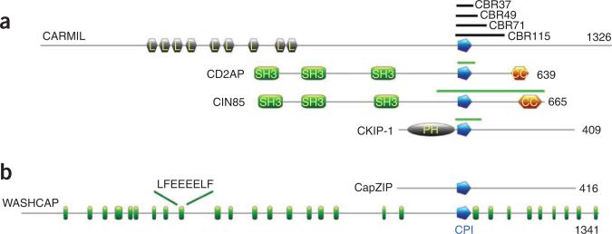
Capping protein (CP) regulates actin dynamics by binding the barbed ends of actin filaments. Removal of CP may be one means to harness actin polymerization for processes such as cell movement and endocytosis. Here we structurally and biochemically investigated a CP interaction (CPI) motif present in the otherwise unrelated proteins CARMIL and CD2AP. The CPI motif wraps around the stalk of the mushroom-shaped CP at a site distant from the actin-binding interface, which lies on the top of the mushroom cap. We propose that the CPI motif may act as an allosteric modulator, restricting CP to a low-affinity, filament-binding conformation. Structure-based sequence alignments extend the CPI motif-containing family to include CIN85, CKIP-1, CapZIP and a relatively uncharacterized protein, WASHCAP (FAM21). Peptides comprising these CPI motifs are able to inhibit CP and to uncap CP-bound actin filaments.


Figures

Figure 1
Figure 1
Domain organization of CPI motif–containing proteins. (a) Previously known CPI family members. L, leucine-rich repeat; SH3, src homology domain 3; PH, pleckstrin homology domain; CC, coiled coil. The green bars above CD2AP, CIN85 and CKIP-1 signify reported CPI regions. Black bars above CARMIL detail the constructs used in this study. CBR37, CBR49, CBR71 and CBR115 comprise residues Ser968–Cys1004, Glu964–Asp1012, Glu964–Val1034 and Glu964–Ser1078, respectively. CBR71 is approximately equivalent to CAH3965–1038 (ref. 10). (b) Novel CPI family members. WASHCAP contains multiple repeats homologous to LF(E/D)nLF (green) that show a high incidence of serine residues. The LF(E/D)nLF motifs are generally followed by basic residue–rich regions.
Figure 2
Figure 2
Functional definition of the CPI motif. (a) Inhibition of CP capping by histidine (His)-tagged CBR115 and CBR37 as monitored by the pyrene-actin polymerization assay. (b) Uncapping of CP-capped actin filaments. Data for His-CBR71 and His-CBR49 are included in Supplementary Figure 2a,b. Pyrene-labeled actin (1.5 μM, 7% pyrene) was polymerized in the presence of spectrin-actin seeds, and actin polymer concentration was monitored by pyrene fluorescence intensity. For the inhibition assay, actin was polymerized in the presence of CP with His-CBR115 or CBR37 at the indicated concentrations (nM). The concentration of CP (chicken α1/β1) was 10 nM for CBR115 and 8 nM for CBR37. For the uncapping assay, actin was polymerized in the presence of CP (10 nM); fragments of CBR were added after 200 s. Actin polymerized in the absence of CP is included as a positive control in these and subsequent pyrene assays. These controls (labeled C) show the fastest polymerization in each experiment. (c) Fluorescence anisotropy analysis of the interaction of Alexa488-CBR37 with CP. (d) Fluorescence anisotropy measurements indicating that the Alexa488-CBR37–CP complex is dissociated upon competition with unlabeled CBR37 but not with G-actin. (e) Fluorescence anisotropy measurements suggesting dissociation of the Alexa488-CBR37–CP complex at high concentrations of F-actin.
Figure 3
Figure 3
CARMIL- and CD2AP-induced uncapping of CP-capped actin filaments monitored by TIRF microscopy. (ac) CBR115 (200 nM) (a), CBR37 (250 μM) (b) and CD2AP CPI peptide (200 μM) (c) support green actin filament growth from the previously capped red actin filaments, indicating uncapping. (d) In the absence of CP, the red actin filaments act as seeds for the green actin. (e) The CP-capped actin filaments do not elongate in the absence of CARMIL or CD2AP truncations. Red actin (2 μM) was polymerized and capped with CP (chicken α1/β1, 50 nM), stabilized with phalloidin and diluted to 50 nM. CARMIL or CD2AP truncations were added to CP-capped red actin filaments concurrently with green actin (0.2 μM) and CP (50 nM). Profilin was maintained throughout at a 1.5 molar ratio over actin. Time stamps (min:sec) for frames are given; “–” refers to before addition.
Figure 4
Figure 4
The structure of the CPΔβTent–CARMIL CPI motif complex. (ac) Three views of the CPΔβTent–CARMIL CPI complex. CARMIL CPI motif is shown as a surface (yellow) and CP α-subunit (red) and β-subunit (blue) as schematic representations. (b) 90° rotation around the x axis with respect to a. (c) 180° rotation around the y axis with respect to a. N and C, N and C termini, respectively. (d) Cartoon representation of a, depicting the mushroom-like CP structure with the CPI motif half-encircling the stalk on the underside of the mushroom cap. The β-tentacle, which was not included in the construct for the structure, is added for completeness. (e) Sequence homology of the CARMIL CPI across species displayed as a sequence logo (http://weblogo.berkeley.edu/) with colors red, blue, yellow and green signifying acidic, basic, hydrophobic and hydrophilic residues, respectively. The major CPI residues, α-subunit (red) and β-subunit (blue), are displayed above the logo. S denotes a residue that plays a structural role; X signifies a noninteracting, nonstructural residue in this complex. A more detailed analysis of the CPΔβTent–CARMIL CPI complex is given in Supplementary Figure 3.
Figure 5
Figure 5
Structural differences between CARMIL and CD2AP in binding CP. (a) The structure of the CPΔβTent–CD2AP CPI motif complex with CARMIL CPI motif structure overlaid. The C-terminal half of the CD2AP CPI motif (cyan) generally follows the path of the CARMIL CPI motif (yellow) in binding the β-subunit of CP. The paths of N termini of the CD2AP and CARMIL CPI motifs differ slightly in binding to the α-subunit. (b) The structure of CBR115–CP. Two ordered CARMIL regions are observed: CPI (Ile971–Cys1004, yellow) and CSI (CARMIL-specific interaction; Arg1021–Thr1035, green). Phenylalanine residues (1029 and 1030) are shown in black. A more comprehensive analysis of the interactions within these structures is given in Supplementary Figure 3.
Figure 6
Figure 6
The CPI motif family of proteins. (a) Structure-based sequence alignment of the CPI motif family. α and β refer to the interacting subunit of CP. (b) Pyrene-actin assays monitoring CP uncapping induced by the six members of the CPI motif family. Concentrations of CP and the 37-mer peptides for CPI motif family are as indicated. Actin monomer and spectrin seeds concentrations are as described for Figure 2. The associated inhibition assays can be found in Supplementary Figure 4e. FAM21 is here referred to as WASHCAP. AU, arbitrary units.
Figure 7
Figure 7
Models of CPI motif uncapping. (a) Model of a CP-capped actin filament. The cyan and orange helices and arrow denote the proposed repositioning of the β-tentacle. The CARMIL CPI (yellow surface) and CSI (green surface) motifs are superimposed on the model. (b) 180° rotation of a around the y axis. The β-tentacle is omitted for clarity. (c) A cartoon depicting a potential uncapping mechanism of β-tentacle sequestration by the CPI motif. (d) A cartoon depicting flexibility in the mushroom cap of CP. (e) Superimposition of two CP structures (α-subunits, red and orange; β-subunits, blue and cyan) showing the relative position of the CPI (yellow surface) and CSI (green surface). The CPI motif is speculated to alter CP flexibility, disfavoring the filament-bound conformation.

References

    1. Kovar DR, Wu JQ, Pollard TD. Profilin-mediated competition between capping protein and formin Cdc12p during cytokinesis in fission yeast. Mol. Biol. Cell. 2005;16:2313–2324. - PMC - PubMed
    1. Wear MA, Yamashita A, Kim K, Maeda Y, Cooper JA. How capping protein binds the barbed end of the actin filament. Curr. Biol. 2003;13:1531–1537. - PubMed
    1. Cooper JA, Sept D. New insights into mechanism and regulation of actin capping protein. Int. Rev. Cell Mol. Biol. 2008;267:183–206. - PMC - PubMed
    1. Mejillano MR, et al. Lamellipodial versus filopodial mode of the actin nanomachinery: pivotal role of the filament barbed end. Cell. 2004;118:363–373. - PubMed
    1. Yamashita A, Maeda K, Maeda Y. Crystal structure of CapZ: structural basis for actin filament barbed end capping. EMBO J. 2003;22:1529–1538. - PMC - PubMed

Publication types