. 2015 Sep 18;290(38):22919-30.
doi: 10.1074/jbc.M115.669333. Epub 2015 Jul 30.

Molecular Insights into Inhibition of the Methylated Histone-Plant Homeodomain Complexes by Calixarenes

Affiliations

Molecular Insights into Inhibition of the Methylated Histone-Plant Homeodomain Complexes by Calixarenes

Muzaffar Ali et al. J Biol Chem. .

Abstract

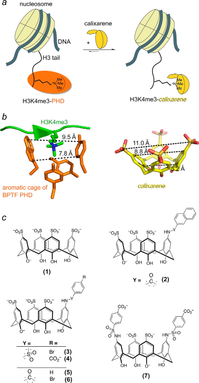
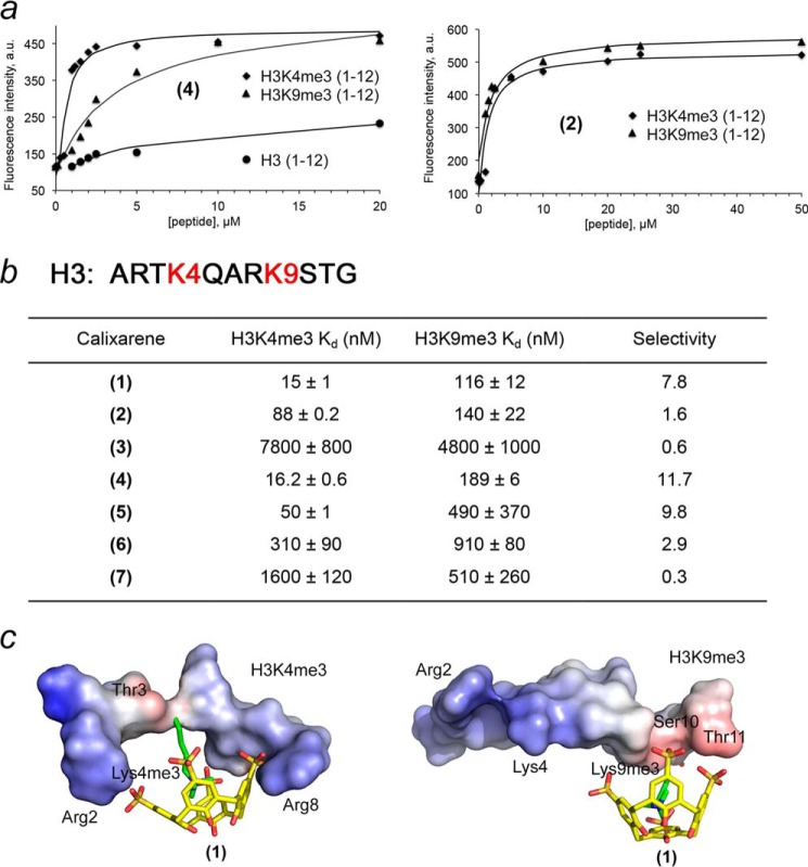
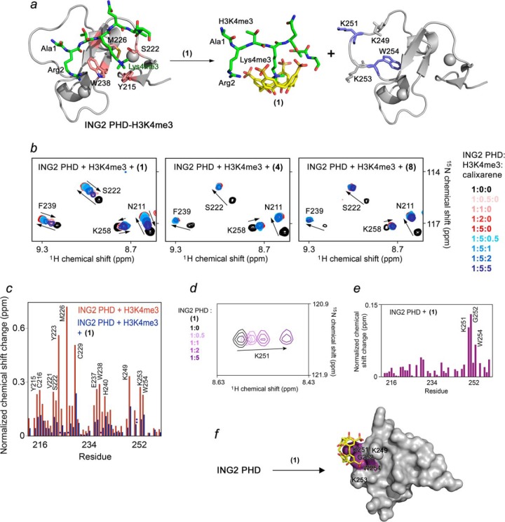
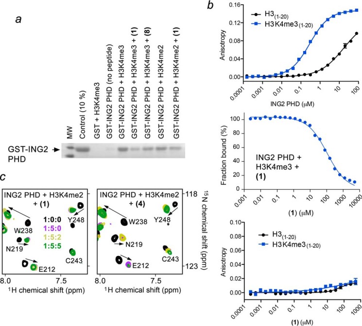
Plant homeodomain (PHD) finger-containing proteins are implicated in fundamental biological processes, including transcriptional activation and repression, DNA damage repair, cell differentiation, and survival. The PHD finger functions as an epigenetic reader that binds to posttranslationally modified or unmodified histone H3 tails, recruiting catalytic writers and erasers and other components of the epigenetic machinery to chromatin. Despite the critical role of the histone-PHD interaction in normal and pathological processes, selective inhibitors of this association have not been well developed. Here we demonstrate that macrocyclic calixarenes can disrupt binding of PHD fingers to methylated lysine 4 of histone H3 in vitro and in vivo. The inhibitory activity relies on differences in binding affinities of the PHD fingers for H3K4me and the methylation state of the histone ligand, whereas the composition of the aromatic H3K4me-binding site of the PHD fingers appears to have no effect. Our approach provides a novel tool for studying the biological roles of methyllysine readers in epigenetic signaling.

Keywords: PHD finger; calixarene; chromatin; histone; histone methylation; inhibitor.


Figures

FIGURE 1.
FIGURE 1.
Calix[4]arenes mimic the aromatic cage of PHD fingers. a, schematic showing the disruption of the association of a PHD finger (orange) with the histone H3K4me3 tail by calixarene (yellow). b, distances between the aromatic groups in the methyllysine-binding cage of the BPTF PHD finger (left panel) and distances between sulfur and carbon atoms in calixarene (right panel) are indicated by dashed lines. c, the calixarene hosts used in this study.
FIGURE 2.
FIGURE 2.
Calixarenes display a selectivity for methylated histone peptides. a, representative FDA binding curves for the indicated peptides and calixarenes. b, dissociation constants measured by FDA. Selectivity for H3K4me3 determined by the ratio of H3K4me3 versus H3K9me3. Data are mean ± S.D. c, modeling of the complexes of (1) with H3K4me3 and H3K9me3 peptides. The electrostatic surface potential of the peptides was generated in PyMOL.
FIGURE 3.
FIGURE 3.
Calixarene (1) inhibits binding of the ING2 PHD finger to H3K4me3. a, molecular model depicting disassociation of the ING2 PHD-H3K4me3 complex because of (1). The crystal structure of the H3K4me3-bound ING2 PHD finger (PDB code 2G6Q) is shown as a ribbon diagram with the H3K4me3 peptide colored green. The ING2 residues comprising the methyllysine-binding aromatic cage are colored pink, whereas residues involved in a nonspecific interaction with (1) are shown in blue. b, superimposed 1H,15N HSQC spectra of ING2 PHD, collected as first H3K4me3 peptide and then (1), (4), or (8) were titrated in. The spectra are color-coded according to the protein:peptide:calixarene molar ratio as shown in the right panel. c, the normalized chemical shift changes observed in the ING2 PHD finger upon binding to H3K4me3 (red) and inhibition by (1) (blue) as a function of residue. *, peaks disappear; **, peaks move in a different direction. d, superimposed 1H,15N HSQC spectra of ING2 PHD collected as (1) was titrated in. e, the normalized chemical shift changes observed in the ING2 PHD finger upon interaction with (1) as a function of residue. f, the residues of the ING2 PHD finger, most perturbed because of the interaction with (1), are mapped onto the structure of the protein.
FIGURE 4.
FIGURE 4.
Calixarene (1) impairs the interaction of the ING2 PHD finger with H3K4me3 but does not disturb the interaction with H3K4me2. a, binding of the GST-fusion ING2 PHD finger to the indicated biotinylated histone peptides in the absence or presence of the indicated calixarenes was examined by pulldown assay. b, quantitation of the interaction of ING2 PHD with H3 (black) or H3K4me3 (blue) peptides by fluorescence polarization (top panel) or fluorescence polarization displacement (center panel) by (1). Bottom panel, quantitation of the interaction of (1) with H3 (black) or H3K4me3 (blue) peptides by fluorescence polarization (bottom). c, overlays of 1H,15N HSQC spectra of the H3K4me2-bound ING2 PHD finger recorded during gradual addition of (1) or (4).
FIGURE 5.
FIGURE 5.
Effects of binding affinity and the aromatic cage topology. a, alignment of PHD finger sequences. Conserved residues are colored pink. The aromatic cage residues of each PHD finger are indicated by purple ovals. b, a close view of the aromatic cages of the PHD fingers of ING2 (PDB code 2G6Q), JARID1A (PDB code 3GL6), MLL1 (PDB code 3LQJ), and MLL5 (PDB code 2LV9) (3, 11, 36, 37). The aromatic cage residues of ING2, JARID1A, MLL1, and MLL5 are labeled and colored pink, yellow, blue, and green, respectively. c, an overlay of the K4me3-binding pockets of ING2, JARID1A, MLL1, and MLL5. d, summary of the Kd values and aromatic cage topologies of the PHD fingers.
FIGURE 6.
FIGURE 6.
Calixarenes exhibit selectivity for the PHD fingers of JARID1A and MLL1. a, the crystal structure of the JARID1A PHD3 finger in complex with H3K4me3 peptide (PDB code 3GL6). The aromatic cage residues are colored yellow. b, superimposed 1H,15N HSQC spectra of JARID1A PHD3, collected as first H3K4me3 peptide and then (1), (4), or (7) were titrated in. The spectra are color-coded according to the protein:peptide:calixarene molar ratio. c, overlays of 1H,15N HSQC spectra of the H3K4me2-bound JARID1A PHD3 recorded during titration with (1). d, superimposed 1H,15N HSQC spectra of MLL1 PHD3 collected as first H3K4me3 peptide and then (1), (4), and (7) were added gradually. e, the crystal structure of the MLL1 PHD3 finger in complex with H3K4me3 peptide (PDB code 3GL6). The aromatic cage residues are colored blue. f, the normalized chemical shift changes observed in the MLL1 PHD finger upon binding to H3K3me3 (red) and inhibition by (1) or (4) (blue) as a function of residue.
FIGURE 7.
FIGURE 7.
Calixarenes block the histone-binding activity of the MLL5 PHD finger in vitro and in vivo. a, crystal structure of the MLL5 PHD finger in complex with H3K4me3 peptide (PDB code 2LV9). The aromatic cage residues are colored green. b, superimposed 1H,15N HSQC spectra of MLL5 PHD, collected as first H3K4me3 peptide and then (1), (4), or (7) were titrated in. The spectra are color-coded according to the protein:peptide:calixarene molar ratio. c, quantitation of the interaction of MLL5 PHD with H3 (black) or H3K4me3 (blue) peptides by fluorescence polarization (left) or fluorescence polarization displacement (right) by (1). d, overlays of 1H,15N HSQC spectra of the H3K4me2-bound MLL5 PHD recorded during titration with (1) or (7). e, binding of the GST-fusion MLL5 PHD finger to the indicated biotinylated histone peptides in the absence or presence of the indicated calixarenes.
FIGURE 8.
FIGURE 8.
Calixarene (1) disrupts binding of MLL5 to H3K4me3 in vivo. a, C2C12 cells expressing FLAG-MLL5 were fixed and stained with anti-FLAG and anti-H3K4me3 antibodies and with secondary antibodies conjugated with Duolink PLA probes and Duolink PLA detection reagents and imaged using a fluorescence microscope. Yellow dots specify interacting foci. Nuclei are labeled blue (DAPI). b, mean ± S.E. of MLL5-H3K4me3-interacting foci per indicated cells. c, percentage of cells with 5–9 (blue) and more than 10 (red) MLL5-H3K4me3-interacting foci. The remaining cells (not shown) contain 0–4 foci. d, levels of FLAG-MLL5 were evaluated by Western blot analysis upon doxycycline (DOX) induction for 24 h. PARP-1 was used as a loading control. e, levels of FLAG-MLL5 were evaluated by immunofluorescence microscopy upon doxycycline induction for 24 h. Identical exposure settings and histogram adjustments were used for each condition (uninduced, left panel, and induced, right panel). f, H3K4me3 levels in C2C12:FLAG-MLL5 cells. Cells were treated with or without 100 μm (1) and processed for immunofluorescence. A z-slice from the nuclear midpoint is shown. Identical exposure settings and histogram adjustments were used for each condition.

References

    1. Musselman C. A., Kutateladze T. G. (2011) Handpicking epigenetic marks with PHD fingers. Nucleic Acids Res. 39, 9061–9071 - PMC - PubMed
    1. Li H., Ilin S., Wang W., Duncan E. M., Wysocka J., Allis C. D., Patel D. J. (2006) Molecular basis for site-specific read-out of histone H3K4me3 by the BPTF PHD finger of NURF. Nature 442, 91–95 - PMC - PubMed
    1. Peña P. V., Davrazou F., Shi X., Walter K. L., Verkhusha V. V., Gozani O., Zhao R., Kutateladze T. G. (2006) Molecular mechanism of histone H3K4me3 recognition by plant homeodomain of ING2. Nature 442, 100–103 - PMC - PubMed
    1. Shi X., Hong T., Walter K. L., Ewalt M., Michishita E., Hung T., Carney D., Peña P., Lan F., Kaadige M. R., Lacoste N., Cayrou C., Davrazou F., Saha A., Cairns B. R., Ayer D. E., Kutateladze T. G., Shi Y., Côté J., Chua K. F., Gozani O. (2006) ING2 PHD domain links histone H3 lysine 4 methylation to active gene repression. Nature 442, 96–99 - PMC - PubMed
    1. Wysocka J., Swigut T., Xiao H., Milne T. A., Kwon S. Y., Landry J., Kauer M., Tackett A. J., Chait B. T., Badenhorst P., Wu C., Allis C. D. (2006) A PHD finger of NURF couples histone H3 lysine 4 trimethylation with chromatin remodelling. Nature 442, 86–90 - PubMed

Publication types

MeSH terms

LinkOut - more resources