. 2015 Dec 18;290(51):30225-39.
doi: 10.1074/jbc.M115.685867. Epub 2015 Oct 16.

Insights into Ubiquitination from the Unique Clamp-like Binding of the RING E3 AO7 to the E2 UbcH5B

Affiliations

Insights into Ubiquitination from the Unique Clamp-like Binding of the RING E3 AO7 to the E2 UbcH5B

Shengjian Li et al. J Biol Chem. .

Abstract

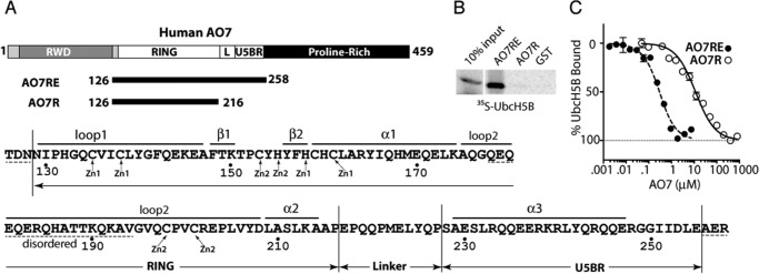
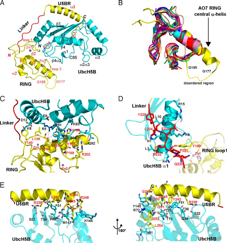
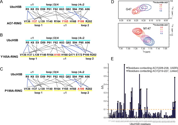
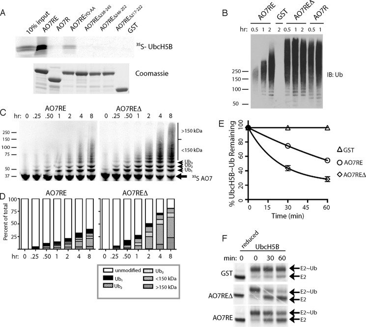
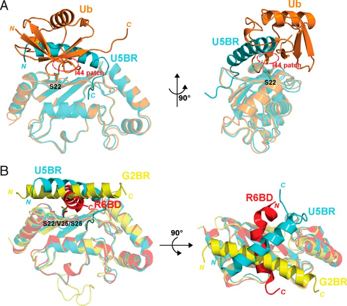
RING proteins constitute the largest class of E3 ubiquitin ligases. Unlike most RINGs, AO7 (RNF25) binds the E2 ubiquitin-conjugating enzyme, UbcH5B (UBE2D2), with strikingly high affinity. We have defined, by co-crystallization, the distinctive means by which AO7 binds UbcH5B. AO7 contains a structurally unique UbcH5B binding region (U5BR) that is connected by an 11-amino acid linker to its RING domain, forming a clamp surrounding the E2. The U5BR interacts extensively with a region of UbcH5B that is distinct from both the active site and the RING-interacting region, referred to as the backside of the E2. An apparent paradox is that the high-affinity binding of the AO7 clamp to UbcH5B, which is dependent on the U5BR, decreases the rate of ubiquitination. We establish that this is a consequence of blocking the stimulatory, non-covalent, binding of ubiquitin to the backside of UbcH5B. Interestingly, when non-covalent backside ubiquitin binding cannot occur, the AO7 clamp now enhances the rate of ubiquitination. The high-affinity binding of the AO7 clamp to UbcH5B has also allowed for the co-crystallization of previously described and functionally important RING mutants at the RING-E2 interface. We show that mutations having marked effects on function only minimally affect the intermolecular interactions between the AO7 RING and UbcH5B, establishing a high degree of complexity in activation through the RING-E2 interface.

Keywords: E3 ubiquitin ligase; RING finger; allosteric regulation; crystallography; proteolysis; ubiquitin; ubiquitin-conjugating enzyme (E2 enzyme).


Figures

FIGURE 1.
FIGURE 1.
A region beyond the AO7 RING finger is required for high-affinity UbcH5B binding. A, schematic of human AO7 (above). Components of the AO7 clamp are indicated in white (L, Linker). Sequence of AO7RE (below) with Zn2+-coordinating residues and structural features noted in “Results”; disordered regions are indicated with dashed lines. B, binding of in vitro transcribed and translated 35S-labeled UbcH5B to N-terminal GST fusions of AO7 prebound to GS. GST fusions were provided in excess relative to UbcH5B, and each was present in equimolar amounts as determined by Coomassie Blue. C, UbcH5B-Green modified with FlAsH was added to increasing amounts of AO7 from which GST had been cleaved. Binding was assessed by MST.
FIGURE 2.
FIGURE 2.
Crystal structure of the AO7RE·UbcH5B complex. A, ribbon diagram of the structure (residue 1 of UbcH5B and residues 126–128, 178–194, and 256–258 of AO7 are not observed and are presumably disordered). UbcH5B is shown in cyan. AO7RE is shown in yellow except for the Linker, in red. Helices, strands, and loops are illustrated as spirals, arrows, and tubes, respectively. The N and C termini and secondary structure elements are labeled in black and red for UbcH5B and AO7, respectively. The disordered peptide (residues 178–194 after the α1 helix) of AO7RE is represented with a dashed curve. Two zinc ions in AO7 RING are illustrated. B, superposition of AO7 RING with other RING and U-box structures (PDB entries 3FL2, 1Z6U, 1FBV, 3HCT, 1LDJ, 2CKL, 2C2V, 3EB6, and 3LRQ). The AO7 RING is shown in yellow, the central α-helix of AO7 RING is labeled, and the disordered peptide in loop 2 of the AO7 RING is indicated with a dashed curve. C–E, the interactions of UbcH5B with AO7 RING (C), Linker (D), and U5BR (E, viewed from two directions). UbcH5B is shown in cyan, RING and U5BR are in yellow, and Linker is in red. The residues involved in electrostatic (cutoff distance = 3.5 Å) and van der Waals (cutoff distance = 4.0 Å) interactions at the interface are shown as sticks. Dashed lines in black indicate salt bridges and hydrogen bonds. The residues involved in the interactions are labeled in black and red for UbcH5B and AO7, respectively.
FIGURE 3.
FIGURE 3.
Interactions between UbcH5B and the U5BR. A–C, summary of intramolecular interactions between WT (A) and mutant (B and C) AO7 RING fingers and UbcH5B. Solid lines indicate van der Waals interactions (cutoff distance = 4.0 Å). Dashed lines indicate hydrogen bonds or salt bridges (cutoff distance = 3.5 Å). The secondary structure assignments of the residues are illustrated. Residues in red were mutated to Ala, and co-crystallization was undertaken or attempted as described in the “Results” section. D, trajectories of two 15N-labeled UbcH5B residues that shift upon titration with AO7 fragment 217–258 (Linker and U5BR) used to calculate Kd. E, chemical shift perturbations in the UbcH5B upon interaction with the AO7 fragment (residues 217–258). Unlabeled AO7 was titrated onto 15N-labeled UbcH5B. Upon titration of 15N-labeled UbcH5B, residues on the UbcH5B backside β-sheet are perturbed significantly in areas corresponding to U5BR binding. In contrast, the Linker does not perturb the UbcH5B significantly. The perturbation (Δδ) is defined as the difference in peak frequencies of the free and AO7-saturated states. Δδ is calculated by the formula Δδ = [(δNf − δNs)2/5 + (δHf − δHs)2]1/2, where δHf, δHs and δNf, δNs are the proton and nitrogen frequencies in free and saturated state, respectively. UbcH5B residues contacting AO7 in the crystal structure are shown as circles on the data. Black circles represent UbcH5B contacts with the AO7 U5BR in the crystal structure, whereas gray-filled circles represent UbcH5B contacts with the AO7 Linker.
FIGURE 4.
FIGURE 4.
Effects of disruption of the U5BR. A, indicated GST fusions of mutations and deletions in AO7RE were assessed for binding to UbcH5B as in Fig. 1B. AO7REYQ-AA refers to Ala mutations of Tyr-242 and Gln-245. B, GST fusions prebound to GS were subject to autoubiquitination reactions and, after washing of beads, eluted material was immunoblotted (IB) for ubiquitin. C, 35S-labeled in vitro transcribed and translated GST fusions were incubated in solution in the presence of E1, UbcH5B, and ubiquitin for the indicated times and ubiquitination assessed. D, quantification of results from C. E, in vitro transcribed and translated 35S-labeled UbcH5B was loaded with ubiquitin. After inactivation of E1, the rate of loss of covalently-bound ubiquitin was measured in the presence of GST-AO7RE or GST-AO7REΔ and ∼80 μm free ubiquitin. Results were normalized to those obtained with GST. Concentrations of GST fusions and E2 were ∼1 μm and 2 nm, respectively. Data represent the average and S.D. of three independent experiments. F, representative experiment used in E.
FIGURE 5.
FIGURE 5.
Role of backside binding in AO7 ubiquitination. A, binding assays were carried out as in Fig. 1B, percent of input bound is indicated. B, time course of ubiquitination was carried out as in Fig. 4B. C, loss of E2∼Ub was assessed as in Fig. 4E. Data are from three independent experiments. D, ubiquitination was carried out as in Fig. 4B. E, binding assay was carried out as in Fig. 1B. IB, immunoblot. F, ubiquitination assay was carried out as in Fig. 4B for 90 min using UbcM2 as the E2. G, purified, bacterially expressed WT or S22R UbcH5B was loaded with equimolar amounts of ubiquitin lacking Lys (UbK0), and the release of UbK0 was assessed during incubation in the presence or absence of added UbK0 (160 μm) and with AO7REΔ from which GST had been cleaved. UbcH5B was detected by immunoblotting. Note the ratio of E2∼Ub to E2 over the time course. Efficiency of loading is less than in experiments using 35S-labeled E2 and WT ubiquitin (e.g. Fig. 4F), as the amount of UbK0 used was kept low during loading to maximize the effect of UbK0 added during the discharge phase.
FIGURE 6.
FIGURE 6.
Assessment of potential for ubiquitin backside binding in cells. A, HEK-293 cells were transfected as indicated with plasmids encoding HA-tagged AO7 amino acids 2–439 (HA-AO7) or HA-AO7 lacking amino acids 238–245 (HA-AO7Δ). Each transfection was divided into four before carrying out a cycloheximide chase. B and C, HEK-293 cells were transfected as in Fig. 5A and treated with either MG132 (50 μm) or lactacystin (LCN; 10 μm). D, HEK-293 cells were transfected with plasmid encoding HA-AO7Δ or HA-AO7Δ in which Tyr-165 in the central α-helix of the RING was mutated to Ala. Cells were then divided for cycloheximide (CHX) chase as in Fig. 6A. GFP served as a transfection efficiency control in all experiments.
FIGURE 7.
FIGURE 7.
Structures of UbcH5B in complex with AO7RE mutants. A, superimposition of the AO7RE·UbcH5B (in cyan), AO7REY165A·UbcH5B (in magenta), and AO7REP199A·UbcH5B (in orange) complexes. B, the Y165A mutation resulted in a change in the AO7 Met-169 side-chain orientation inasmuch as the Met-169 side chain partially occupies the space created by the Y165A mutation, causing the AO7 Glu-172 side chain to bend toward the Met-169 side chain such that Glu-172 forms a new salt bridge with Lys-63 in the β3β4 loop of UbcH5B. The His-168 side chain, which is disordered in the WT, is stabilized in the mutant. Such conformational changes reshape the local E2-binding surface of the RING. Furthermore, several water molecules (W1, W2, and W3) invade into the cavity created by the Y165A mutation. The mutant and WT structures are shown in magenta and cyan, respectively. Residues at the RING-E2 interface are shown as sticks. The water molecules are shown as spheres. C, the P199A mutation brings a water molecule (W1) into the hydrophobic core of AO7 RING, which is hydrogen-bonded to the main chain amide of the Ala-199 residue. This does not disturb the conformation of nearby side chains. The residues on the interface or surrounding the hydrophobic core and the water molecule are illustrated. The mutant and WT structures are shown in orange and cyan, respectively.
FIGURE 8.
FIGURE 8.
Consequences of RING finger mutations. A, 35S-labeled UbcH5B was incubated with the indicated GST fusions as in Fig. 1B. B, data were from MST as in Fig. 1C, including the same data set for WT AO7RE. See Table 1 for calculated the Kd values. C, equal amounts of the indicated AO7RE mutations were purified in complex with UbcH5B by washing co-expressed complexes only once, and ubiquitination was assessed after the addition of an equal amount of purified UbcH5B. Twice as much E2 was added in the GST control reaction. A Coomassie stain of the purified complexes is shown below. D, single round turnover experiments were carried out as in Fig. 4E. Data are graphed as the fraction of E2∼Ub remaining at each time point and are the average of three independent experiments.
FIGURE 9.
FIGURE 9.
Backside binding to E2s. A, comparison of the AO7RE U5BR·UbcH5B (in cyan) and non-covalently associated ubiquitin (UbB)·UbcH5B (in orange) complexes. The superposition is based on the UbcH5B Cα positions. The Ser-22 side chain of UbcH5B is shown as sticks, and the Ile-44 hydrophobic patch of ubiquitin is circled in red. The U5BR helix crosses the loop connecting the β3 and β4 strands of ubiquitin. Ser-22 of UbcH5B is also covered by the overlapping region of the U5BR and ubiquitin. B, comparison of the AO7RE U5BR·UbcH5B (in cyan), gp78 G2BR·Ube2g2 (in magenta) and Rad18 R6BD·Rad6B (in red) complexes. The superposition of the complexes is based on the E2 Cα positions. Ser-22 in UbcH5B and the corresponding residues in Ube2G2 and Rad6B (Val-25 and Ser-25, respectively) are shown as sticks. G2BR contains only a straight helix and lies on the backside of E2; R6BD is a twisted helix and spans the β-sheet surface of Rad6b. The U5BR has an extra C-terminal loop in addition to the long helix, anchors to a hydrophobic valley on the UbcH5B surface, and thus provides more extensive interaction with E2. The indicated N and C termini are of ubiquitin, U5BR, G2BR and R6BD.

References

    1. Metzger M. B., Pruneda J. N., Klevit R. E., and Weissman A. M. (2014) RING-type E3 ligases: master manipulators of E2 ubiquitin-conjugating enzymes and ubiquitination. Biochim. Biophys. Acta 1843, 47–60 - PMC - PubMed
    1. Deshaies R. J., and Joazeiro C. A. (2009) RING domain E3 ubiquitin ligases. Annu. Rev. Biochem. 78, 399–434 - PubMed
    1. Lorick K. L., Jensen J. P., Fang S., Ong A. M., Hatakeyama S., and Weissman A. M. (1999) RING fingers mediate ubiquitin-conjugating enzyme (E2)-dependent ubiquitination. Proc. Natl. Acad. Sci. U.S.A. 96, 11364–11369 - PMC - PubMed
    1. Weissman A. M., Shabek N., and Ciechanover A. (2011) The predator becomes the prey: regulating the ubiquitin system by ubiquitylation and degradation. Nat. Rev. Mol. Cell Biol. 12, 605–620 - PMC - PubMed
    1. Kirisako T., Kamei K., Murata S., Kato M., Fukumoto H., Kanie M., Sano S., Tokunaga F., Tanaka K., and Iwai K. (2006) A ubiquitin ligase complex assembles linear polyubiquitin chains. EMBO J. 25, 4877–4887 - PMC - PubMed

Publication types

MeSH terms

Substances