. 2011 Feb 8;108(6):2414-9.
doi: 10.1073/pnas.1015433108. Epub 2011 Jan 24.

Crystal structure of a gammadelta T-cell receptor specific for the human MHC class I homolog MICA

Affiliations

Crystal structure of a gammadelta T-cell receptor specific for the human MHC class I homolog MICA

Bin Xu et al. Proc Natl Acad Sci U S A. .

Abstract

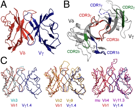
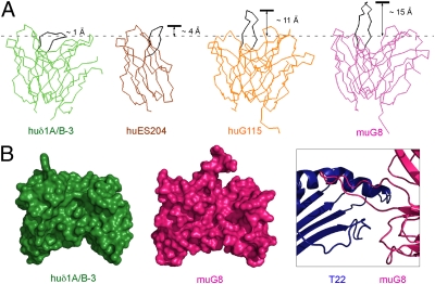
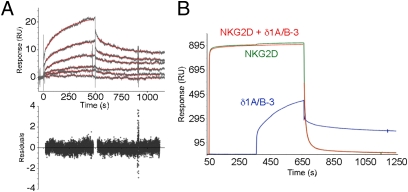
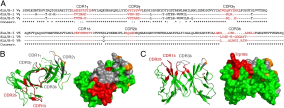
γδ T cells play important roles in bridging innate and adaptive immunity, but their recognition mechanisms remain poorly understood. Human γδ T cells of the V(δ)1 subset predominate in intestinal epithelia and respond to MICA and MICB (MHC class I chain-related, A and B; MIC) self-antigens, mediating responses to tumorigenesis or viral infection. The crystal structure of an MIC-reactive V(δ)1 γδ T-cell receptor (TCR) showed expected overall structural homology to antibodies, αβ, and other γδ TCRs, but complementary determining region conformations and conservation of V(δ)1 use revealed an uncharacteristically flat potential binding surface. MIC, likewise, serves as a ligand for the activating immunoreceptor natural killer group 2, D (NKG2D), also expressed on γδ T cells. Although MIC recognition drives both the TCR-dependent stimulatory and NKG2D-dependent costimulatory signals necessary for activation, interaction analyses showed that MIC binding by the two receptors was mutually exclusive. Analysis of relative binding kinetics suggested sequential recognition, defining constraints for the temporal organization of γδ T-cell/target cell interfaces.


Conflict of interest statement

The authors declare no conflict of interest.

Figures

Fig. 1.
Fig. 1.
The overall structure of an MIC-reactive δ1A/B-3 and comparison with other γδ TCR structures. (A and B) Structure of δ1A/B-3 is shown in two orientations, viewing from the side (A) or top (CDR side; B). In B, the color codes are green for CDR1s (residues 26–35 for CDR1γ and 160–169 for CDR1δ), blue for CDR2s (residues 53–59 for CDR2γ and 185–189 for CDR2δ), red for CDR3s (residues 98–106 for CDR3γ and 228–239 for CDR3δ), and light gray for the rest of the molecule. Parts of the CDR1γ and HV4 loops that were not modeled are depicted as dotted lines. (C) Superposition of human Vδ2Vγ9 G115, Vδ3 δ-chain ES204, and murine G8 γδ TCRs onto human Vδ1 δ1A/B-3. Color codes are red for Vδ, blue or light gray for Vγ, orange for Vδ2, cyan for Vδ3 δ-chain, and magenta for G8.
Fig. 2.
Fig. 2.
Comparison of γδ TCR CDR3δ loop conformations. (A) Three previously determined γδ TCR structures are superimposed onto the δ1A/B-3 structure and displayed in identical orientations. Color (and PDB codes) are green for δ1A/B-3, orange for G115 (1HXM), magenta for murine G8 (1YPZ), and brown for Vδ3 δ-chain ES204 (1TVD). CDR3δ loops are all highlighted in black. (B) Variable domains of human Vδ1 and murine G8 are displayed in the same orientation in a space-filling representation. The protrusion of CDR3δ in G8 relative to δ1A/B-3 is evident. Part of the G8:T22 complex is shown in Right, highlighting the role of CDR3δ in T22 recognition.
Fig. 3.
Fig. 3.
SPR kinetic analysis of δ1A/B-3:MICA and competitive binding in δ1A/B-3:NKG2D:MICA interactions. (A) SPR sensorgrams of δ1A/B-3:MICA binding are shown, with TCR analyte concentrations of 5, 2.5, 1, 0.5, 0.25, and 0.1 μM. Fitted binding curves are shown as red traces, and corresponding fitting residuals are shown below. (B) SPR-based analysis of the competition between NKG2D and δ1A/B-3 for binding to MICA. Sensorgrams from different channels are colored as blue for δ1A/B-3, green for NKG2D alone, and red for NKG2D (first 5 min) and NKG2D plus δ1A/B-3 (after the first 5 min).
Fig. 4.
Fig. 4.
MIC-reactive Vδ1 γδ TCR sequences and CDR surface conservation mapped onto the δ1A/B-3 structure. (A) Amino acid sequences of three MIC-responsive Vδ1 γδ TCR clones are shown, designated as δ1A/B-1, δ1A/B-3, and δ1A/B-5 (10), with CDR loops highlighted in red. (B and C) Two views of the δ1A/B-3 structure are shown as ribbons (Left) or molecular surfaces (Right). Identical CDR residues are colored red, identical framework residues are colored green, conservatively substituted residues are colored orange throughout, and nonconserved residues are colored gray throughout.

References

    1. Hayday AC. Gammadelta T cells and the lymphoid stress-surveillance response. Immunity. 2009;31:184–196. - PubMed
    1. Hayday AC. [gamma][delta] cells: A right time and a right place for a conserved third way of protection. Annu Rev Immunol. 2000;18:975–1026. - PubMed
    1. Boismenu R, Havran WL. An innate view of gamma delta T cells. Curr Opin Immunol. 1997;9:57–63. - PubMed
    1. Chien YH, Konigshofer Y. Antigen recognition by gammadelta T cells. Immunol Rev. 2007;215:46–58. - PubMed
    1. Rudolph MG, Stanfield RL, Wilson IA. How TCRs bind MHCs, peptides, and coreceptors. Annu Rev Immunol. 2006;24:419–466. - PubMed

Publication types

MeSH terms

Substances

Associated data

LinkOut - more resources