. 2011 May 1;67(Pt 5):541-5.
doi: 10.1107/S1744309111010761. Epub 2011 Apr 20.

Structure of Mycobacterium tuberculosis phosphopantetheine adenylyltransferase in complex with the feedback inhibitor CoA reveals only one active-site conformation

Affiliations

Structure of Mycobacterium tuberculosis phosphopantetheine adenylyltransferase in complex with the feedback inhibitor CoA reveals only one active-site conformation

T Wubben et al. Acta Crystallogr Sect F Struct Biol Cryst Commun. .

Abstract

Phosphopantetheine adenylyltransferase (PPAT) catalyzes the penultimate step in the coenzyme A (CoA) biosynthetic pathway, reversibly transferring an adenylyl group from ATP to 4'-phosphopantetheine to form dephosphocoenzyme A (dPCoA). To complement recent biochemical and structural studies on Mycobacterium tuberculosis PPAT (MtPPAT) and to provide further insight into the feedback regulation of MtPPAT by CoA, the X-ray crystal structure of the MtPPAT enzyme in complex with CoA was determined to 2.11 Å resolution. Unlike previous X-ray crystal structures of PPAT-CoA complexes from other bacteria, which showed two distinct CoA conformations bound to the active site, only one conformation of CoA is observed in the MtPPAT-CoA complex.


Figures
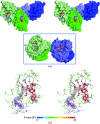
Figure 1
Figure 1
MtPPAT in complex with CoA. (a) Wall-eyed stereoview of the asymmetric unit of the MtPPAT–CoA crystal complex. Two protomers belong to one hexamer (shades of green) and the other two belong to an adjacent hexamer (shades of blue) in the crystal (inset). The two protomers from each respective hexamer are arranged across the dyad axis. The protomers are shown in surface representation. CoA is bound to all four protomers in the asymmetric unit and is depicted in space-filling representation. C atoms are shown in gray, N atoms in blue, S atoms in yellow, O atoms in red and phosphates in orange. (b) Wall-eyed stereoview of the amino-acid residues and water molecules in contact with CoA in the active site of MtPPAT. Subunit A of MtPPAT is colored forest green. Residues from the adjacent subunit across the trimer threefold axis are shown in slate and marked with an asterisk. Hydrogen bonds are depicted as dashed lines and water molecules are depicted as red spheres. CoA is colored according to the B factor of each atom and the F oF c electron density (light magenta) of CoA is contoured at 2.5σ, while the 2F oF c electron density (gray) is contoured at 1.0σ. The mean B factor of the protein is 25.0 Å2, whereas the mean B factor of all four CoA molecules in the asymmetric unit is 50.6 Å2 (49.2 Å2 for the CoA molecule in subunit A). The mean B factor of CoA is greater than that of the protein, mainly owing to the disorder of the CoA adenylate moiety.
Figure 2
Figure 2
Relationship between the different modes of ligand binding to MtPPAT. (a) Superposition of different ligand-complexed forms of MtPPAT. Subunit A of MtPPAT–CoA is shown in green, MtPPAT–PhP is shown in orange (PDB code 3nbk, subunit A; Wubben & Mesecar, 2010 ▶) and MtPPAT–AMPcPP is shown in blue (PDB code 3nba, subunit C; Wubben & Mesecar, 2010 ▶). CoA (magenta), PhP (gray) and AMPcPP (cyan) are shown in ball-and-stick representation. (b) Projection of the adenylate moiety of CoA into the solvent channel of the biologically significant hexamer of MtPPAT. The view is down the triad axis of the hexamer. MtPPAT (green) is depicted in cartoon representation, with CoA shown in ball-and-stick representation.
Figure 3
Figure 3
Comparison of the CoA-binding mode in PPAT from different organisms. Y. pestis PPAT–CoA (PDB code 3l92, magenta; unpublished work), E. coli PPAT–CoA subunit A (PDB code 1h1t, orange; Izard, 2003 ▶) and E. coli PPAT–CoA subunit B (PDB code 1h1t, blue; Izard, 2003 ▶) are superimposed onto MtPPAT–CoA (subunit A, forest green). CoA bound to Y. pestis PPAT is shown in hot pink, CoA bound to E. coli PPAT is shown in yellow (subunit A) and blue (subunit B) and CoA bound to MtPPAT is shown in green. Residues from MtPPAT, E. coli PPAT subunit B and Y. pestis PPAT that are marked with an asterisk are from the adjacent subunit across the triad axis. Residues from subunit A of E. coli PPAT were not labeled because they are the same as those labeled from subunit B.

References

    1. Bruzzese, F. J. & Connelly, P. R. (1997). Biochemistry, 36, 10428–10438. - PubMed
    1. Chen, V. B., Arendall, W. B., Headd, J. J., Keedy, D. A., Immormino, R. M., Kapral, G. J., Murray, L. W., Richardson, J. S. & Richardson, D. C. (2010). Acta Cryst. D66, 12–21. - PMC - PubMed
    1. Edwards, T. E., Leibly, D. J., Bhandari, J., Statnekov, J. B., Phan, I., Dieterich, S. H., Abendroth, J., Staker, B. L., Van Voorhis, W. C., Myler, P. J. & Stewart, L. J. (2011). Acta Cryst. F67, doi:10.1107/S1744309111004349. - PMC - PubMed
    1. Fernández-Millán, P., Kortazar, D., Lucas, M., Martínez-Chantar, M. L., Astigarraga, E., Fernández, J. A., Sabas, O., Albert, A., Mato, J. M. & Martínez-Cruz, L. A. (2008). Acta Cryst. F64, 605–609. - PMC - PubMed
    1. Geerlof, A., Lewendon, A. & Shaw, W. V. (1999). J. Biol. Chem. 274, 27105–27111. - PubMed

Publication types

MeSH terms

Associated data