. 2011 Feb 25;331(6020):1036-40.
doi: 10.1126/science.1195380. Epub 2010 Dec 16.

Structure of DNMT1-DNA complex reveals a role for autoinhibition in maintenance DNA methylation

Affiliations

Structure of DNMT1-DNA complex reveals a role for autoinhibition in maintenance DNA methylation

Jikui Song et al. Science. .

Abstract

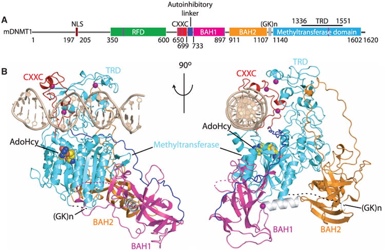
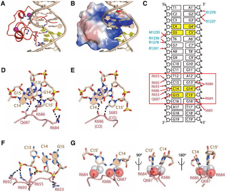
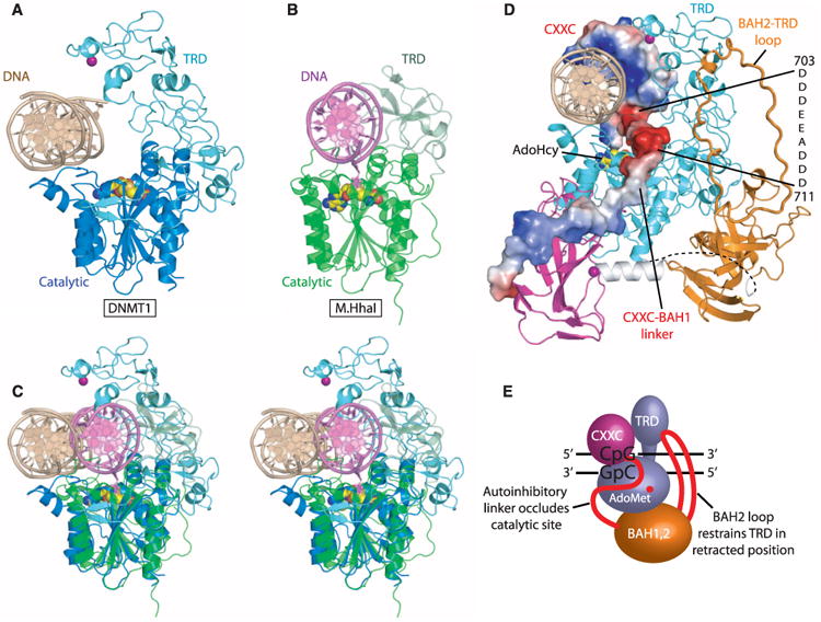
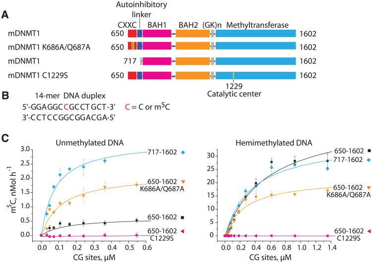
Maintenance of genomic methylation patterns is mediated primarily by DNA methyltransferase-1 (DNMT1). We have solved structures of mouse and human DNMT1 composed of CXXC, tandem bromo-adjacent homology (BAH1/2), and methyltransferase domains bound to DNA-containing unmethylated CpG sites. The CXXC specifically binds to unmethylated CpG dinucleotide and positions the CXXC-BAH1 linker between the DNA and the active site of DNMT1, preventing de novo methylation. In addition, a loop projecting from BAH2 interacts with the target recognition domain (TRD) of the methyltransferase, stabilizing the TRD in a retracted position and preventing it from inserting into the DNA major groove. Our studies identify an autoinhibitory mechanism, in which unmethylated CpG dinucleotides are occluded from the active site to ensure that only hemimethylated CpG dinucleotides undergo methylation.


Figures

Fig. 1
Fig. 1
Structural overview of mDNMT1(650–1602)–DNA 19-nucleotide oligomer complex with bound AdoHcy. (A) Color-coded domain architecture and numbering of mDNMT1 sequence. The thin vertical light blue bars indicate binding positions of zinc ions. (B) Ribbon representation of the complex in two orthogonal views. The CXXC, BAH1, BAH2, and methyltransferase domain are colored in red, light purple, orange and light blue, and DNA and zinc ions are colored in light brown and dark purple, respectively; CXXC-BAH1 linker in dark blue, BAH1-BAH2 linker in silver, (GK)n-containing BAH2-methyltransferase linker in black, and bound AdoHcy as in space-filling representation.
Fig. 2
Fig. 2
Intermolecular contacts between CXXC domain of mDNMT1(650–1602) and DNA 19-nucleotide oligomer. (A) Ribbon representation of the CXXC domain bound to DNA. The CpG step is rendered in yellow. (B) Surface electrostatic representation of the CXXC domain bound to DNA. (C) Schematic view of intermolecular interactions involving the CXXC domain in the mDNMT1-DNA 19-nucleotide oligomer complex (boxed red rectangle), with intermolecular contacts shown by red arrows. The residue labels are colored according to their respective domains in Fig. 1. (D) Hydrogen-bonding interactions between side chains from the CXXC domain and the guanine base edges of the CpG step in the DNA major groove. The nitrogen, oxygen, and phosphorous atoms are shown in dark blue, red, and yellow, respectively. The bases of the CpG dinucleotide from one strand are shaded. (E) Hydrogen-bonding interactions between backbone carbonyl oxygens from the CXXC domain and cytosine amino groups of the CpG step in the DNA major groove. (F) Hydrogen-bonding interactions between arginine side chains of the CXXC domain and the phosphodiester backbone of the DNA along the DNA minor groove. (G) Potential steric clashes between methylated cytosine modeled on either strand of the CpG step and the CXXC domain of DNMT1 in the structure of the complex. Van der Waals radii are rendered in red for DNMT1 and gray for modeled methylated cytosine.
Fig. 3
Fig. 3
Comparison of mDNMT1 with M.HhaI in their DNA-bound complexes. (A) The crystal structure of the mDNMT1(650–1602)–DNA 19-nucleotide oligomer complex. The CXXC, BAH1, and BAH2 domains and CXXC-BAH1 linker of mDNMT1 have been removed for clarity. The bound DNA is in light brown, with the TRD and catalytic core in light and dark blue, respectively. (B) The crystal structure of the M.HhaI-DNA complex [PDB: 1MHT (8)]. The bound DNA is in light purple, with the TRD and catalytic core in pale and dark green, respectively. (C) Structural comparison of mDNMT1(650–1602)–DNA 19-nucleotide oligomer complex and M.HhaI-DNA complex in a stereo view looking down the DNA helix axis, after superposition of their methyltransferase domains. AdoHcy is shown in space-filling view. The everted cytosine in the M.HhaI complex is shown in ball-and-stick view in dark purple. (D) Electrostatic surface representation of mDNMT1 CXXC domain and the CXXC-BAH1 linker in the context of the structure of the mDNMT1(650–1602)–DNA 19-nucleotide oligomer complex. The BAH2-TRD loop is highlighted with thicker lines. (E) The proposed model for autoinhibitory mechanism in maintenance DNA methylation. In the autoinhibitory state, the CXXC domain and the auto-inhibitory linker (in red) occlude the active site. In addition, the BAH2-TRD loop (in red) restrains the TRD in a retracted position so that it does not interact with CpG sites on the DNA.
Fig. 4
Fig. 4
Efficiency of unmethylated and hemimethylated DNA m5C-methylation by mouse wild-type and mutant DNMT1 proteins. (A) Domain structures of mouse DNMT1 proteins used for activity assay. The Lys686Ala/Gln687Ala and Cys1229Ser mutations are shown as yellow bars. (B) Sequence of 14-nucleotide oligomer DNA duplex used for enzymatic assay. (C) Enzyme kinetics on de novo and maintenance methylation of DNMT1 mutants after removal or mutation of CXXC domain. Rates of 3H-CH3 in nMol per hour transferred to unmethylated and hemimethylated DNA duplexes by 1 nM of mDNMT1(650–1602) wild-type, Lys686Ala/Gln687 Ala and Cys1229Ser mutants and mDNMT1(717–1602) proteins were plotted as a function of CpG site concentration; the steady-state Michaelis-Menten parameters that were estimated from these plots are listed in table S3. Each reaction point was repeated in triplicate; mean and SD values are shown.

Comment in

References

    1. Law JA, Jacobsen SE. Nat Rev Genet. 2010;11:204. - PMC - PubMed
    1. Cheng X, Blumenthal RM. Structure. 2008;16:341. - PMC - PubMed
    1. Goll MG, Bestor TH. Annu Rev Biochem. 2005;74:481. - PubMed
    1. Li E, Beard C, Jaenisch R. Nature. 1993;366:362. - PubMed
    1. Bourc'his D, Bestor TH. Nature. 2004;431:96. - PubMed

Publication types

MeSH terms