. 2011 Dec 2;414(3):356-69.
doi: 10.1016/j.jmb.2011.09.047. Epub 2011 Oct 12.

Structure of the Escherichia coli phosphonate binding protein PhnD and rationally optimized phosphonate biosensors

Affiliations

Structure of the Escherichia coli phosphonate binding protein PhnD and rationally optimized phosphonate biosensors

Ismael Alicea et al. J Mol Biol. .

Abstract

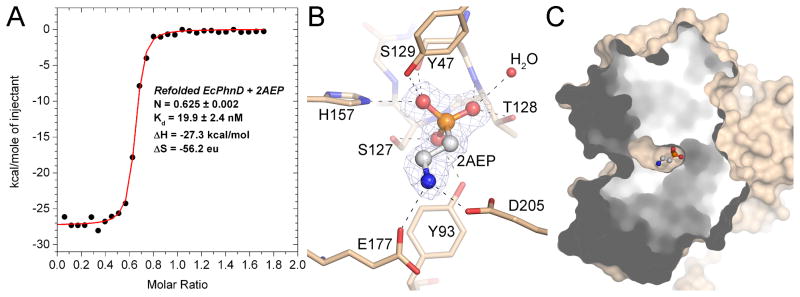
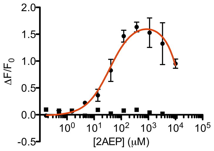
The phnD gene of Escherichia coli encodes the periplasmic binding protein of the phosphonate (Pn) uptake and utilization pathway. We have crystallized and determined structures of E. coli PhnD (EcPhnD) in the absence of ligand and in complex with the environmentally abundant 2-aminoethylphosphonate (2AEP). Similar to other bacterial periplasmic binding proteins, 2AEP binds near the center of mass of EcPhnD in a cleft formed between two lobes. Comparison of the open, unliganded structure with the closed 2AEP-bound structure shows that the two lobes pivot around a hinge by ~70° between the two states. Extensive hydrogen bonding and electrostatic interactions stabilize 2AEP, which binds to EcPhnD with low nanomolar affinity. These structures provide insight into Pn uptake by bacteria and facilitated the rational design of high signal-to-noise Pn biosensors based on both coupled small-molecule dyes and autocatalytic fluorescent proteins.


Figures

FIGURE 1
FIGURE 1. Binding of 2AEP to EcPhnD
A, ITC measurement of 2AEP binding to EcPhnD, following denaturing, washing, and refolding. Binding parameters calculated from the best fit binding model are inset. B, The phosphonate-binding site of EcPhnD with 2AEP bound. Selected portions of EcPhnD are displayed as sticks with tan color carbons. 2AEP is shown as ball-and-stick with white carbons. A portion of the 2FoFc electron density omit map for 2AEP contoured at 2σ is shown as grey mesh. C, The solvent accessible surface of EcPhnD is shown with the front portion cut away to show the buried ligand binding pocket containing 2AEP (ball-and-stick).
FIGURE 2
FIGURE 2. Overall structure of EcPhnD
A, Representation of the primary structure of EcPhnD colored by structural domain. B, Cartoon representation of the crystal structure of EcPhnD bound to 2AEP colored by domain as in A. 2AEP is shown as ball-and-stick. C, Topology diagram of the EcPhnD structure, colored as in A and B. α-helices are represented as circles, β-strands as triangles. Darker shaded shapes represent secondary structure elements that are not part of the conserved type II PBP fold. D, E, Cartoon representations of the closed, 2AEP-bound (tan) and open (burgundy) structures of EcPhnD, superimposed on Lobe 1 (panel D) or on Lobe 2 (panel E). A two-headed arrow points to equivalent regions of the structures, illustrating the magnitude of conformational change.
FIGURE 3
FIGURE 3. Oligomeric state of EcPhnD
A, SEC chromatograms of EcPhnD and C-terminal truncations. A calibration curve produced using protein standards is inset. Molecular weights estimated from each peak elution volume are shown as color coded dots and labels superimposed on the inset calibration curve. Theoretical molecular weights of each species are given in parentheses. B, Cartoon representation of the dimerization interface between two EcPhnD molecules (colored tan and blue). Selected side-chains participating in the dimerization interface are shown as sticks. 2AEP is shown as ball-and-stick. M304 of the tan molecule is labeled to highlight the site of the smallest truncation that disrupted dimerization.
FIGURE 4
FIGURE 4. In vitro screening for exogenous-fluorophore phosphonate sensors
A, Change in fluorescence from IANBD-conjugated EcPhnD single cysteine variants following addition of either buffer control (black bars) or 1 mM 2AEP (red bars). Fluorescence titrations of purified, IANBD-conjugated EcPhnD variants Q17C (B) and K181C (C) with 2AEP were then carried out. Curve fit was a single-site binding isotherm [y=((Fmax−Fo)/(1+(Kd/x)))+Fo]. Error bars show standard deviation (n = 3).
FIGURE 5
FIGURE 5. A genetically encoded phosphonate sensor
A, Fluorescence titration of the best genetically encoded phosphonate sensor, EcPhnD90-cpGFP.L1ADΔΔ.L297R,L301R, with 2AEP. Titration of a cpGFP fusion with a glucose binding protein that has no affinity for phosphonates is shown in square symbols as a negative control. We attribute the marked decrease of fluorescence at high concentrations of 2AEP in the sensor titration to fluorescence quenching. Curve fit was a single-site binding isotherm plus a linear component to account for quenching. Error bars show standard deviation (n = 3).

References

    1. Wanner BL. In: Escherichia coli and Salmonella: cellular and molecular biology 2nd edit. Neidhardt FC, Curtiss R III, Ingraham JL, Lin ECC, Low KB, Magasanik B, Reznikoff W, Riley M, Schaechter M, Umbarger HE, editors. ASM Press; Washington, D.C.: 1996. pp. 1357–1381.
    1. Metcalf WW, Wanner BL. Mutational analysis of an Escherichia coli fourteen-gene operon for phosphonate degradation, using TnphoA’ elements. J Bacteriol. 1993;175:3430–42. - PMC - PubMed
    1. Gebhard S, Cook GM. Differential regulation of high-affinity phosphate transport systems of Mycobacterium smegmatis: identification of PhnF, a repressor of the phnDCE operon. J Bacteriol. 2008;190:1335–43. - PMC - PubMed
    1. Jochimsen B, Lolle S, McSorley FR, Nabi M, Stougaard J, Zechel DL, Hove-Jensen B. Five phosphonate operon gene products as components of a multi-subunit complex of the carbon-phosphorus lyase pathway. Proc Natl Acad Sci USA 2011 - PMC - PubMed
    1. Hove-Jensen B, Rosenkrantz TJ, Haldimann A, Wanner BL. Escherichia coli phnN, encoding ribose 1,5-bisphosphokinase activity (phosphoribosyl diphosphate forming): dual role in phosphonate degradation and NAD biosynthesis pathways. J Bacteriol. 2003;185:2793–801. - PMC - PubMed

Publication types

MeSH terms

LinkOut - more resources