. 2011 Jul 8;286(27):23900-10.
doi: 10.1074/jbc.M111.242016. Epub 2011 May 11.

Crystal structures of Staphylococcus epidermidis mevalonate diphosphate decarboxylase bound to inhibitory analogs reveal new insight into substrate binding and catalysis

Affiliations

Crystal structures of Staphylococcus epidermidis mevalonate diphosphate decarboxylase bound to inhibitory analogs reveal new insight into substrate binding and catalysis

Michael L Barta et al. J Biol Chem. .

Abstract

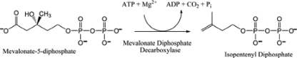
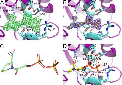
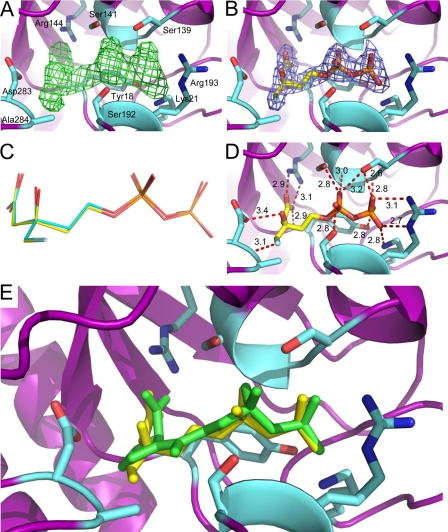
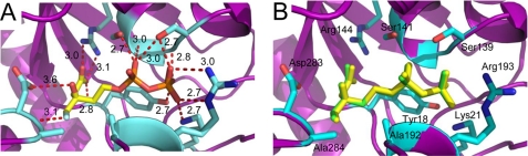
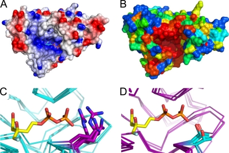
The polyisoprenoid compound undecaprenyl phosphate is required for biosynthesis of cell wall peptidoglycans in gram-positive bacteria, including pathogenic Enterococcus, Streptococcus, and Staphylococcus spp. In these organisms, the mevalonate pathway is used to produce the precursor isoprenoid, isopentenyl 5-diphosphate. Mevalonate diphosphate decarboxylase (MDD) catalyzes formation of isopentenyl 5-diphosphate in an ATP-dependent irreversible reaction and is therefore an attractive target for inhibitor development that could lead to new antimicrobial agents. To facilitate exploration of this possibility, we report the crystal structure of Staphylococcus epidermidis MDD (1.85 Å resolution) and, to the best of our knowledge, the first structures of liganded MDD. These structures include MDD bound to the mevalonate 5-diphosphate analogs diphosphoglycolyl proline (2.05 Å resolution) and 6-fluoromevalonate diphosphate (FMVAPP; 2.2 Å resolution). Comparison of these structures provides a physical basis for the significant differences in K(i) values observed for these inhibitors. Inspection of enzyme/inhibitor structures identified the side chain of invariant Ser(192) as making potential contributions to catalysis. Significantly, Ser → Ala substitution of this side chain decreases k(cat) by ∼10(3)-fold, even though binding interactions between FMVAPP and this mutant are similar to those observed with wild type MDD, as judged by the 2.1 Å cocrystal structure of S192A with FMVAPP. Comparison of microbial MDD structures with those of mammalian counterparts reveals potential targets at the active site periphery that may be exploited to selectively target the microbial enzymes. These studies provide a structural basis for previous observations regarding the MDD mechanism and inform future work toward rational inhibitor design.


Figures

REACTION 1
REACTION 1
FIGURE 1.
FIGURE 1.
Inhibition of S. epidermidis MDD by MVAPP analogs. Double-reciprocal plots of MDD activity versus substrate MVAPP concentration are shown as measured in the presence of either DPGP or FMVAPP. A, inhibition of MDD by DPGP. Concentrations of DPGP are as follows: 0 μm (●), 3 μm (○), 5 μm (▾), 7 μm (▿), and 10 μm (■). B, inhibition of MDD by FMVAPP. Concentrations of FMVAPP are as follows: 0 nm (●), 40 nm (○), 80 nm (▾), 160 nm (▿), and 320 nm (■). Concentrations of substrate (MVAPP) were 2.5, 5, 10, 20, and 40 μm in both panels. Optimal fits to the data were obtained using a mixed inhibition model in A and a competitive inhibition model in B.
FIGURE 2.
FIGURE 2.
1.85 Å crystal structure of apo-MDD from S. epidermidis. A, crystal structure of S. epidermidis MDD shown in ribbon format. Two copies of MDD are found within the asymmetric unit and are colored blue (N terminus) and red (C terminus). B, stereo view of MDD structures in ribbon format deposited within the PDB. Structures correspond to the following organisms: S. epidermidis (red-orange); S. aureus (cyan); L. pneumophila (orange); S. pyogenes (red); H. sapiens (yellow-green); S. cerevisiae (blue); T. brucei (green); and M. musculus (yellow). C, limited structure-based sequence alignment of prokaryotic and eukaryotic MDD proteins. Alignment was generated using ClustalW and rendered with ESPRIPT. Numbers above the sequences correspond to S. epidermidis MDD. Red stars below the sequences correspond to invariant amino acid side chains involved in DPGP and FMVAPP interaction, and the green star represents the single variable active site residue. Sequences from B as well as the following were used in alignment: L. monocytogenes; E. faecalis; Xenopus. tropicalis; B. taurus; A. thaliana.
FIGURE 3.
FIGURE 3.
2.05 Å cocrystal structure of MDD bound to the inhibitor DPGP. A, FoFc map (green mesh at 2.0σ contour) of the refined structure in the absence of modeled ligand. Active site side chains within interaction distance are depicted in ball and stick format (cyan). MDD backbone is depicted in ribbon format (purple). B, 2FoFc map (blue mesh at 2.0σ contour) of the refined structure with one molecule DPGP modeled per enzyme. Color scheme is the same as A, with DPGP colored yellow. C, overlay of DPGP molecules from the active sites of chains A (yellow) and B (cyan). D, active site side chains within 2.6–3.4 Å of DPGP. Further information on these distances can be found in supplemental Table S1.
FIGURE 4.
FIGURE 4.
2.20 Å cocrystal structure of MDD bound to the inhibitor FMVAPP. A, FoFc map (green mesh at 3.0σ contour) of the refined structure in the absence of modeled ligand. Active site side chains within interaction distance are depicted in ball and stick format (cyan). MDD backbone is depicted in ribbon format (purple). B, 2FoFc map (blue mesh at 2.0σ contour) of the refined structure with one molecule FMVAPP modeled per enzyme. Color scheme is the same as A, with FMVAPP colored yellow. C, overlay of FMVAPP molecules from chains A (yellow) and B (cyan). D, active site side chains within 2.6–3.4 Å of FMVAPP. Further information on these distances can be found in supplemental Table S2. E, overlay of the MDD active site from cocrystal structures of DPGP (green) and FMVAPP (yellow). Active site side chains from the FMVAPP cocrystal structure are depicted in ball and stick format (cyan).
FIGURE 5.
FIGURE 5.
2.10 Å cocrystal structure of MDD mutant S192A bound to the inhibitor FMVAPP. A, active site side chains within 2.6–3.6Å of FMVAPP are depicted in ball and stick format (cyan). Further information on these distances can be found in supplemental Table S2. MDD backbone is depicted in ribbon format (purple). B, overlay of FMVAPP molecules from wild type MDD (green) and mutant S192A MDD (yellow). Color scheme is the same as A.
FIGURE 6.
FIGURE 6.
Conserved nature of the active site cleft. A, surface representation of electrostatic potential, generated by DelPhi, of chain A from MDD bound to FMVAPP (cyan). Color scheme represents regions of negative (red) and positive (blue) charge density contoured at ±5 e/kT. B, conservation plot of surface-exposed MDD amino acid side chains from S. epidermidis, as generated by CONSURF. Color scheme depicts variable (blue to green), average (yellow), and strong (orange to red) conservation. C, conserved nature of Arg193 (purple stick) within the active site of prokaryotic MDD proteins (backbone atoms colored cyan). Superimposed MDD structures are from S. epidermidis, S. aureus, S. pyogenes, and L. pneumophila. D, conserved nature of Thr (cyan stick) in eukaryotic MDD proteins (backbone atoms in purple) at the structurally equivalent position to Arg193. Superimposed MDD structures are from H. sapiens, M. musculus, T. brucei, and S. cerevisiae. FMVAPP is shown in C and D according to its position in the wild type MDD + FMVAPP structure (Fig. 4).

References

    1. Zetola N., Francis J. S., Nuermberger E. L., Bishai W. R. (2005) Lancet Infect. Dis. 5, 275–286 - PubMed
    1. Tenover F. C., Biddle J. W., Lancaster M. V. (2001) Emerging Infect. Dis. 7, 327–332 - PMC - PubMed
    1. Lowy F. D. (1998) N. Engl. J. Med. 339, 520–532 - PubMed
    1. Eisenreich W., Bacher A., Arigoni D., Rohdich F. (2004) Cell. Mol. Life Sci. 61, 1401–1426 - PMC - PubMed
    1. Wilding E. I., Brown J. R., Bryant A. P., Chalker A. F., Holmes D. J., Ingraham K. A., Iordanescu S., So C. Y., Rosenberg M., Gwynn M. N. (2000) J. Bacteriol. 182, 4319–4327 - PMC - PubMed

Publication types

MeSH terms