. 2011 Jun 17;286(24):21295-303.
doi: 10.1074/jbc.M111.226282. Epub 2011 Apr 19.

Structural basis for microcin C7 inactivation by the MccE acetyltransferase

Affiliations

Structural basis for microcin C7 inactivation by the MccE acetyltransferase

Vinayak Agarwal et al. J Biol Chem. .

Abstract

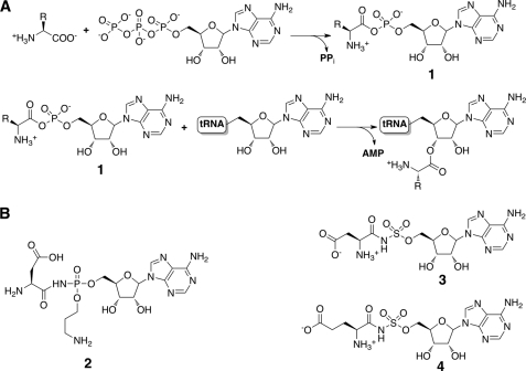
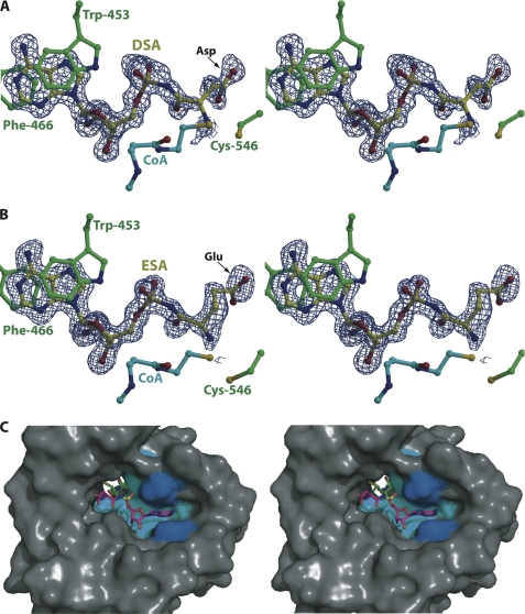
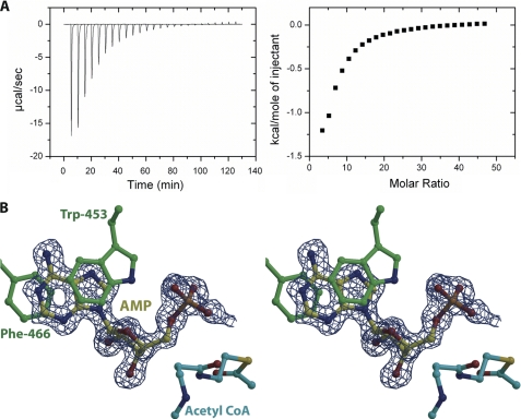
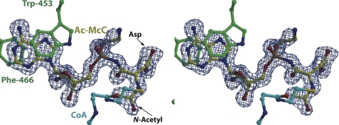
The antibiotic microcin C7 (McC) acts as a bacteriocide by inhibiting aspartyl-tRNA synthetase and stalling the protein translation machinery. McC is synthesized as a heptapeptide-nucleotide conjugate, which is processed by cellular peptidases within target strains to yield the biologically active compound. As unwanted processing of intact McC can result in self-toxicity, producing strains utilize multiple mechanisms for autoimmunity against processed McC. We have shown previously that the mccE gene within the biosynthetic cluster can inactivate processed McC by acetylating the antibiotic. Here, we present the characterization of this acetylation mechanism through biochemical and structural biological studies of the MccE acetyltransferase domain (MccE(AcTase)). We have also determined five crystal structures of the MccE-acetyl-CoA complex with bound substrates, inhibitor, and reaction product. The structural data reveal an unexpected mode of substrate recognition through π-stacking interactions similar to those found in cap-binding proteins and nucleotidyltransferases. These studies provide a rationale for the observation that MccE(AcTase) can detoxify a range of aminoacylnucleotides, including those that are structurally distinct from microcin C7.


Figures

FIGURE 1.
FIGURE 1.
tRNA synthetase mechanism and structures of inhibitors. A, the two-step reaction for the esterification of a specific amino acid onto its cognate tRNA to form a charged aminoacyl-tRNA proceeds through the production of a hydrolytically labile aminoacyl adenylate (compound 1). B, chemical structures of synthetic and naturally occurring aminoacyl adenylate mimics, including McC (compound 2), DSA (compound 3), and ESA (compound 4).
FIGURE 2.
FIGURE 2.
Overall structure and multiple sequence alignment. A, ribbon diagram showing the overall structure of MccEAcTase (in cyan) in complex with acetyl-CoA (yellow ball and stick). B, close-up view of the acetyl-CoA-binding site. Superimposed is a difference Fourier electron density map (in blue) calculated with coefficients |Fobs| − |Fcalc| and phases from the final refined model with the coordinates of acetyl-CoA deleted prior to one round of refinement. The map is contoured at 3σ over background. C, a structure-based sequence alignment of MccEAcTase along with the GNAT acetyltransferases RimL and YdeF. The active site general acid/base are colored in red, the cysteine residue that is disulfide-bonded to CoA is shown in cyan, and Trp-453 and Phe-466, which are involved in π-stacking interactions with the substrate, are shown in blue. S. enterica, Salmonella enterica.
FIGURE 3.
FIGURE 3.
Active site of MccEAcTase in complex with CoA and DSA/ESA. A, the DSA substrate is colored in yellow, the CoA molecule is colored in cyan, and polypeptide residues colored in green include Trp-453 and Phe-466 that form a π-stacked sandwich with the adenine ring of the substrate and Cys-546 that is disulfide-bonded to the CoA thiol. Superimposed is a difference Fourier electron density map (contoured at 3σ over background and shown in blue) calculated with coefficients |Fobs| − |Fcalc| and phases from the final refined model with the coordinates of DSA deleted prior to one round of refinement. B, the ESA substrate and protein residues are colored as above, and the superimposed difference Fourier electron density map (contoured at 3σ over background and shown in blue) is calculated as above. C, stereoview of a surface rendering of the substrate-binding hydrophobic pocket of MccEAcTase with the superimposed coordinates of DSA (in magenta) and CoA (in green). The hydrophobic cluster of residues (Ile-440, Met-451, Val-493, and Trp-511) that are in van der Waals contact with the substrate is colored in cyan, and Trp-453 and Phe-466 that form a π-stacking interaction with the adenine ring of the substrate are colored in blue.
FIGURE 4.
FIGURE 4.
AMP acts as competitive inhibitor. A, raw data and corresponding binding isotherm for the interaction of MccEAcTase with AMP. B, stereoview of the active site of MccEAcTase in complex with CoA and AMP. Substrate and protein residues are colored and the superimposed difference Fourier electron density map (contoured at 3σ over background and shown in blue) was calculated as in Fig. 3.
FIGURE 5.
FIGURE 5.
Co-crystal structure with acetylated McC product. Stereoview of the active site of MccEAcTase in complex with CoA and acetylated McC (colored as in Fig. 3) with a superimposed difference Fourier electron density map (contoured at 3σ over background and shown in blue) calculated with coefficients |Fobs| − |Fcalc| and phases from the final refined model with the coordinates of substrate deleted prior to one round of refinement.

References

    1. Destoumieux-Garzón D., Peduzzi J., Rebuffat S. (2002) Biochimie 84, 511–519 - PubMed
    1. Hurdle J. G., O'Neill A. J., Chopra I. (2005) Antimicrob. Agents Chemother. 49, 4821–4833 - PMC - PubMed
    1. Pohlmann J., Brötz-Oesterhelt H. (2004) Curr. Drug Targets Infect. Disord. 4, 261–272 - PubMed
    1. Severinov K., Semenova E., Kazakov A., Kazakov T., Gelfand M. S. (2007) Mol. Microbiol. 65, 1380–1394 - PubMed
    1. Guijarro J. I., González-Pastor J. E., Baleux F., San Millán J. L., Castilla M. A., Rico M., Moreno F., Delepierre M. (1995) J. Biol. Chem. 270, 23520–23532 - PubMed

Publication types

MeSH terms

LinkOut - more resources