. 2011 Jun 2;474(7349):54-60.
doi: 10.1038/nature10139. Epub 2011 May 15.

Principles of activation and permeation in an anion-selective Cys-loop receptor

Affiliations

Principles of activation and permeation in an anion-selective Cys-loop receptor

Ryan E Hibbs et al. Nature. .

Abstract

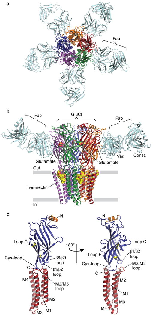
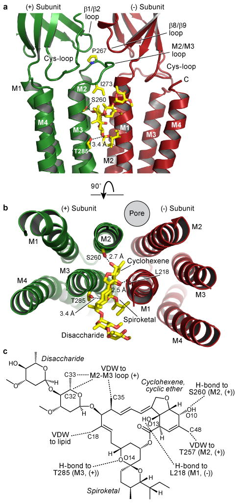
Fast inhibitory neurotransmission is essential for nervous system function and is mediated by binding of inhibitory neurotransmitters to receptors of the Cys-loop family embedded in the membranes of neurons. Neurotransmitter binding triggers a conformational change in the receptor, opening an intrinsic chloride channel and thereby dampening neuronal excitability. Here we present the first three-dimensional structure, to our knowledge, of an inhibitory anion-selective Cys-loop receptor, the homopentameric Caenorhabditis elegans glutamate-gated chloride channel α (GluCl), at 3.3 Å resolution. The X-ray structure of the GluCl-Fab complex was determined with the allosteric agonist ivermectin and in additional structures with the endogenous neurotransmitter L-glutamate and the open-channel blocker picrotoxin. Ivermectin, used to treat river blindness, binds in the transmembrane domain of the receptor and stabilizes an open-pore conformation. Glutamate binds in the classical agonist site at subunit interfaces, and picrotoxin directly occludes the pore near its cytosolic base. GluCl provides a framework for understanding mechanisms of fast inhibitory neurotransmission and allosteric modulation of Cys-loop receptors.


Figures

Figure 1
Figure 1. Architecture of GluClcryst-Fab complex
In a, view of the GluClcryst-Fab complex looking down pore axis toward cytosol. Fab molecules (cyan) are bound at each GluClcryst subunit interface. In b, view parallel to lipid membrane; only two Fab molecules are shown for clarity. The ligands ivermectin and glutamate are represented as spheres with carbon atoms in yellow, oxygen in red and nitrogen in blue. In c, a single GluClcryst subunit from two angles, approximate orientation as in panel b. The Cys-loop and loop C disulfide bonds are shown as spheres, N- and C-termini and TM spans are indicated. Loops of particular relevance to agonist binding and allosteric gating linkage are also indicated.
Figure 2
Figure 2. Ivermectin binding site and atomic interactions
In a and b, two orientations of a GluCl subunit interface focusing on ivermectin binding site. Dashed lines indicate hydrogen bonds. In a, view is from receptor periphery looking parallel to the membrane, and in b looking down pore from extracellular side with ECD removed for clarity. In c, chemical structure of ivermectin with interactions indicated; VDW: van der Waals. Atomic numbering is from PDB file.
Figure 3
Figure 3. Glutamate binding site and specificity
In a, view from extracellular side toward membrane at glutamate in binding site in subunit interface. In b, view of binding site looking parallel to membrane with loop C removed for clarity. Dashed lines with distances in Å indicate hydrogen bonding and, in the case of Tyr200, cation-π interactions. Unless a range is given, distances are an average from the five binding sites. In c, radioligand competition experiments with L-glutamate and congeners against 1 mM [3H]-L-glutamate. Calculated KI values assume a KD for [3H]-L-glutamate of 680 nM and are shown in inset table; n = 2 and CI, confidence interval. L-HCS and L-AA are L-homocysteine sulfinic acid and L-amino adipic acid, respectively.
Figure 4
Figure 4. Ion channel
In a, purple spheres represent internal surface of transmembrane ion channel, with side chains shown for pore-lining residues from two of the five M2 α-helices that line the pore; Ser260 does not line the pore but hydrogen bonds with ivermectin. In b, pore diameter is plotted as a function of longitudinal distance along the pore for GluClcryst, open (GLIC, PDB: 3EAM) and closed (ELIC: 2VL0) bacterial receptors.
Figure 5
Figure 5. Picrotoxin binding site
In a, front two subunits removed to show picrotoxin location (boxed) at cytosolic base of pore. Residues involved in picrotoxin binding are shown as sticks and van der Waals surfaces are shown for picrotoxin. In b, looking into pore from ECD at picrotoxin position relative to 2′Thr and -2′Pro side chains.
Figure 6
Figure 6. Ion selectivity
In a, front of receptor is cut away to reveal interior surface of pore, colored by electrostatic potential. Dashed circle in pore indicates putative chloride binding site. In b, expanded view from a showing selected M2 side chains from opposing subunits. Anomalous difference peaks at pore base are attributed to iodide binding sites (light grey mesh, contoured at 3.5 σ). Fobs-Fcalc omit density for the putative chloride site is represented by yellow mesh contoured at 3 σ. The electropositive pockets where iodide ions bind are shown in c, viewed from inside the protein surface; four residues from adjacent M2 helices that coordinate the iodide sites are shown as sticks. In d, electropositive pockets viewed from the intracellular side. In e, putative chloride site viewed from extracellular side of pore with 6′Thr residues in foreground; Fobs-Fcalc omit density (yellow) is contoured at 4 σ. Carbon atoms are colored by chain and chloride is represented by a 1 Å cyan sphere. Closest distances in Å from protein atoms to center of sphere are indicated by dashed lines from the 6′Thr side chain hydroxyl.

References

    1. Coombs JS, Eccles JC, Fatt P. The specific ionic conductances and the ionic movements across the motoneuronal membrane that produce the inhibitory post-synaptic potential. J Physiol. 1955;130:326–374. - PMC - PubMed
    1. Hille B. Ion Channels of Excitable Membranes. Sinauer Associates, Inc.; Sunderland, MA: 2001.
    1. Thompson AJ, Lester HA, Lummis SC. The structural basis of function in Cys-loop receptors. Q Rev Biophys. 2010;43:449–499. - PubMed
    1. Corringer PJ, et al. Atomic structure and dynamics of pentameric ligand-gated ion channels: new insight from bacterial homologues. J Physiol. 2010;588:565–572. - PMC - PubMed
    1. Hilf RJ, Dutzler R. A prokaryotic perspective on pentameric ligand-gated ion channel structure. Curr Opin Struct Biol. 2009;19:418–424. - PubMed

Publication types

MeSH terms