

Revisiting the role of glycosylation in the structure of human IgG Fc
- PMID: 22747430
- PMCID: PMC3448853
- DOI: 10.1021/cb300130k
Revisiting the role of glycosylation in the structure of human IgG Fc
Abstract
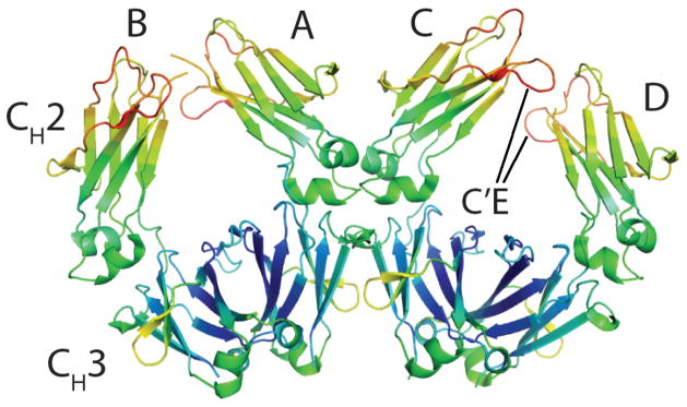
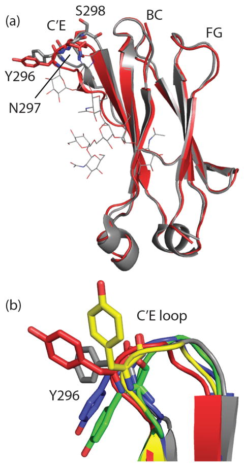
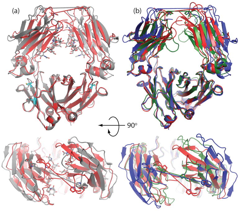
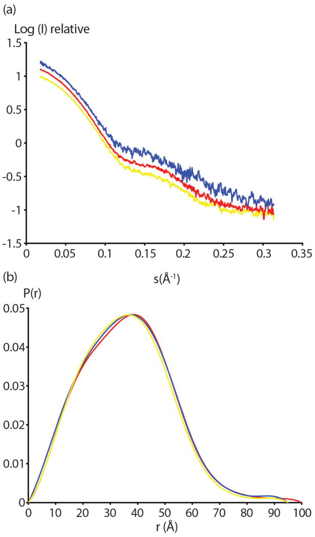
Binding of the Fc domain of Immunoglobulin G (IgG) to Fcγ receptors on leukocytes can initiate a series of signaling events resulting in antibody-dependent cell-mediated cytotoxicity (ADCC) and other important immune responses. Fc domains lacking glycosylation at N297 have greatly diminished Fcγ receptor binding and lack the ability to initiate a robust ADCC response. Earlier structural studies of Fc domains with either full length or truncated N297 glycans led to the proposal that these glycans can stabilize an "open" Fc conformation recognized by Fcγ receptors. We determined the structure of an E. coli expressed, aglycosylated human Fc domain at 3.1 Å resolution and observed significant disorder in the C'E loop, a region critical for Fcγ receptor binding, as well as a decrease in distance between the C(H)2 domains relative to glycosylated Fc structures. However, comparison of the aglycosylated human Fc structure with enzymatically deglycosylated Fc structures revealed large differences in the relative orientations and distances between C(H)2 domains. To provide a better appreciation of the physiologically relevant conformation of the Fc domain in solution, we determined Radii of Gyration (R(g)) by small-angle X-ray scattering (SAXS) and found that the aglycosylated Fc displays a larger R(g) than glycosylated Fc, suggesting a more open C(H)2 orientation under these conditions. Moreover, the R(g) of aglycosylated Fc was reduced by mutations at the C(H)2-C(H)3 interface (E382V/M428I), which confer highly selective binding to FcγRI and novel biological activities.
Conflict of interest statement
The authors declare not conflict of interest
Figures
References
-
- Nimmerjahn F, Ravetch J. Antibodies, Fc receptors and cancer. Curr Opin Immunol. 2007;19:239–245. - PubMed
-
- Nimmerjahn F, Ravetch J. Fc receptors as regulators of immune responses. Nat Rev Immunol. 2008;8:34–47. - PubMed
-
- Jefferis R, Lund J, Pound JD. IgG-Fc-mediated effector functions: molecular definition of interaction sites for effector ligands and the role of glycosylation. Immunol Rev. 1998;163:59–76. - PubMed
-
- Jefferis R. Glycoforms of human IgG in health and disease. Trends in Glycosci Glyc. 2009;21:105–117.
-
- Nesspor TC, Raju TS, Chin CN, Vafa O, Brezski RJ. Avidity confers FcgR binding and immune effector function to aglycosylated immunoglobulin G1. J Mol Recognit. 2012;25:147–154. - PubMed
Publication types
MeSH terms
Substances
Associated data
- Actions
Grants and funding
LinkOut - more resources
Full Text Sources
Other Literature Sources
