. 2015 Feb;89(4):2024-40.
doi: 10.1128/JVI.02968-14. Epub 2014 Nov 26.

Human noroviruses' fondness for histo-blood group antigens

Affiliations

Human noroviruses' fondness for histo-blood group antigens

Bishal K Singh et al. J Virol. 2015 Feb.

Abstract

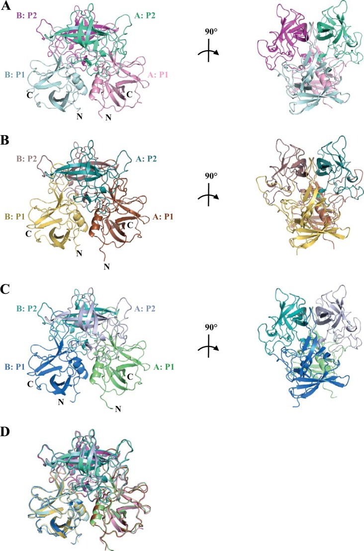
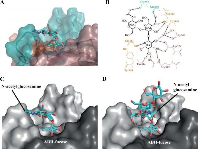
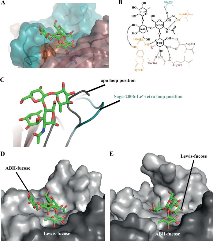
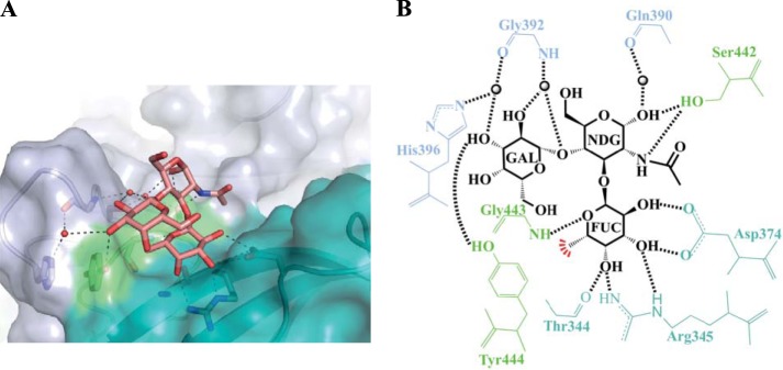
Human noroviruses are the dominant cause of outbreaks of gastroenteritis around the world. Human noroviruses interact with the polymorphic human histo-blood group antigens (HBGAs), and this interaction is thought to be important for infection. Indeed, synthetic HBGAs or HBGA-expressing enteric bacteria were shown to enhance norovirus infection in B cells. A number of studies have found a possible relationship between HBGA type and norovirus susceptibility. The genogroup II, genotype 4 (GII.4) noroviruses are the dominant cluster, evolve every other year, and are thought to modify their binding interactions with different HBGA types. Here we show high-resolution X-ray crystal structures of the capsid protruding (P) domains from epidemic GII.4 variants from 2004, 2006, and 2012, cocrystallized with a panel of HBGA types (H type 2, Lewis Y, Lewis B, Lewis A, Lewis X, A type, and B type). Many of the HBGA binding interactions were found to be complex, involving capsid loop movements, alternative HBGA conformations, and HBGA rotations. We showed that a loop (residues 391 to 395) was elegantly repositioned to allow for Lewis Y binding. This loop was also slightly shifted to provide direct hydrogen- and water-mediated bonds with Lewis B. We considered that the flexible loop modulated Lewis HBGA binding. The GII.4 noroviruses have dominated outbreaks over the past decade, which may be explained by their exquisite HBGA binding mechanisms, their fondness for Lewis HBGAs, and their temporal amino acid modifications.

Importance: Our data provide a comprehensive picture of GII.4 P domain and HBGA binding interactions. The exceptionally high resolutions of our X-ray crystal structures allowed us to accurately recognize novel GII.4 P domain interactions with numerous HBGA types. We showed that the GII.4 P domain-HBGA interactions involved complex binding mechanisms that were not previously observed in norovirus structural studies. Many of the GII.4 P domain-HBGA interactions we identified were negative in earlier enzyme-linked immunosorbent assay (ELISA)-based studies. Altogether, our data show that the GII.4 norovirus P domains can accommodate numerous HBGA types.


Figures

FIG 1
FIG 1
Amino acid alignment of norovirus GII.4 variants. The P domain amino acid sequences of four GII.4 variants, from 1998, 2004, 2006, and 2012 (termed VA387-1998, Farm-2004, Saga-2006, and NSW-2012, respectively), were aligned using Clustal X. The capsid sequences shared 93 to 95% amino acid identity. The S domain was highly conserved, with only seven amino acid differences (not shown), whereas the P1 (red) and P2 (yellow) subdomains were more variable. The common set of amino acids interacting with HBGAs is shaded in blue (chain A) and green (chain B). Compared to the sequence of the earlier discovered GII.4 variant P domain (VA387-1998), one amino acid insertion was observed in 2004 and remained in 2006 and 2012.
FIG 2
FIG 2
X-ray crystal structures of unbound GII.4 P domains. (A) The Farm-2004 P domain apo structure contained one dimer per asymmetric unit. The P domain was subdivided into P1 (chain A in pink and chain B in pale cyan) and P2 (chain A in green cyan and chain B in light magenta) subdomains. (B) The Saga-2006 P domain apo structure contained one monomer per asymmetric unit (a dimer is shown) and was subdivided into P1 (chain A in brown and chain B in yellow-orange) and P2 (chain A in deep teal and chain B in dirty violet) subdomains. (C) The NSW-2012 P domain apo structure contained one monomer per asymmetric unit (a dimer is shown) and was subdivided into P1 (chain A in lime and chain B in marine) and P2 (chain A in blue-white and chain B in teal) subdomains. (D) Superposition of the Farm-2004, Saga-2006, and NSW-2012 P dimers revealed that their overall structures were similar.
FIG 3
FIG 3
Amino acid variations in GII.4 P dimers from 2004, 2006, and 2012 variants. Amino acid changes (red) were highlighted on GII.4 P dimers (side and top views). The changes were numbered according to a change from 1998 to the respective year (labeled once). A cumulative addition of amino acid changes was found. (A) Farm-2004 contained a single amino acid insertion, Gly394, and this remained in 2012. A small number of amino acid changes surrounding the HBGA pocket (black circle), i.e., I389V, L375F, and Q376E, was observed. (B) Saga-2006 contained additional changes, several of which became fixed, e.g., L375F and Q376E. (C) NSW-2012 showed the majority of changes, including several changes in the P1 subdomain.
FIG 4
FIG 4
Representative simulated annealing difference omit maps. The omit maps (blue) were contoured between 2.5 and 2.0 σ. H2-tri is an α-l-fucose-(1-2)-β-d-galactose-(1-4)-N-acetyl-β/α-d-glucosamine, A-tri is an α-l-fucose-(1-2)-β-d-galactose-(3-1)-N-acetyl-α-d-galactosamine, B-tri is an α-l-fucose-(1-2)-β/α-d-galactose-(3-1)-α-d-galactose, Ley-tetra is an α-l-fucose-(1-2)-β-d-galactose-(1-4)-N-acetyl-β/α-d-glucosamine-(3-1)-α-l-fucose, Leb-tetra is an α-l-fucose-(1-2)-β-d-galactose-(1-3)-N-acetyl-β-d-glucosamine-(4-1)-α-l-fucose, Lea-tri is a β-d-galactose-(1-3)-N-acetyl-β/α-d-glucosamine-(4-1)-α-l-fucose (the electron density was well defined for fucose and partially defined for the N-acetylglucosamine and galactose), and Lex-tri is a β-d-galactose-(1-4)-N-acetyl-β/α-d-glucosamine-(3-1)-α-l-fucose. Underlining represents the position of the reducing end hydroxyl group, which was fixed in the α position in the crystal structures.
FIG 5
FIG 5
Saga-2006 P dimer binding interactions with H2-tri. (A) Closeup surface and ribbon representation of the Saga-2006–H2-tri complex structure, showing the hydrogen bonds (black lines) with H2-tri (cyan sticks) and water-mediated interactions (red spheres). (B) Saga-2006 P dimer and H2-tri binding interactions. FUC, α-fucose; GAL, β-galactose; NDG, α-N-acetylglucosamine. The black lines represent the hydrogen bonds, the red line represents the hydrophobic interaction with the hydroxyl group of Tyr444, and the sphere represents water. (C) The ABH fucose of Saga-2006–H2-tri bound at the regular pocket. The acetyl group of N-acetylglucosamine was leaning toward the edge of the P dimer. (D) The ABH fucose of TCH05-2004–H1-pentasaccharide bound at the regular pocket. The acetyl group of N-glucosamine was leaning toward the center of the P dimer.
FIG 6
FIG 6
Saga-2006 P dimer binding interactions with Ley-tetra. (A) Closeup surface and ribbon representation of the Saga-2006–Ley-tetra complex structure, showing the hydrogen bonds with Ley-tetra (green sticks) and water-mediated interactions. (B) Saga-2006 P dimer and Ley-tetra binding interactions. FUC, α-fucose; NDG, α-N-acetylglucosamine; GAL, β-galactose. (C) A loop in the Saga-2006 P2 subdomain (residues 391 to 394) was repositioned from the apo position (gray) to an alternative position (deep teal) for Ley-tetra binding. (D) The Lewis fucose of Saga-2004–Ley-tetra bound at the regular pocket on the P domain and was leaning toward the edge of the P dimer. (E) The ABH fucose of GII.10 P domain Ley-tetra bound at the regular pocket and was orientated toward the center of the P dimer.
FIG 7
FIG 7
Farm-2004 and Saga-2006 P dimer binding interactions with Leb-tetra. (A) Closeup surface and ribbon representation of the Farm-2004–Leb-tetra complex structure, showing the hydrogen bonds with Leb-tetra (marine sticks) and water-mediated interactions. (B) Farm-2004 P dimer and Leb-tetra binding interactions. FUC, α-fucose; GAL, β-galactose; NAG, β-N-acetylglucosamine. The black lines represent the hydrogen bonds, the red line represents the hydrophobic interaction with the hydroxyl group of Tyr444, and the sphere represents water. (C) Closeup surface and ribbon representation of the Saga-2006–Leb-tetra complex structure, showing the hydrogen bonds with Leb-tetra and water-mediated interactions. (D) Saga-2006 P dimer and Leb-tetra binding interactions. (E) Superposition of both the A and B chains of the Farm-2004 apo (gray and black) and Farm-2004–Leb-tetra (cyan and pink) structures.
FIG 8
FIG 8
Saga-2006 P dimer binding interactions with Lea-tri and superposition of GII.4 P domains. (A) Closeup surface and ribbon representation of the Saga-2006–Lea-tri complex structure, showing hydrogen bonds with Lea-tri (camel-colored sticks) and water-mediated interactions. (B) Saga-2006 P dimer and Lea-tri binding interactions. FUC, α-fucose; NDG, α-N-acetylglucosamine; GAL, β-galactose. (C) Superposition of apo and HBGA-bound Farm-2004, Saga-2006, and NSW-2012 P dimer structures (with HBGAs removed from the structures). The circles represent the HBGA binding pocket. Farm-2004 P1 subdomains (chain A in pink and chain B in pale cyan) and P2 subdomains (chain A in green cyan and chain B in light magenta), Saga-2006 P1 subdomains (chain A in brown and chain B in yellow-orange) and P2 subdomains (chain A in deep teal and chain B in dirty violet), and NSW-2012 P1 subdomains (chain A in lime and chain B in marine) and P2 subdomains (chain A in blue-white and chain B in teal) are indicated by color coding. (D) Close-up of the P2 subdomain flexible loop (residues 391 to 394). In the case of H2-tri, Ley-tetra, Lea-tri, and Lex-tri, the N-acetylglucosamine was held by the side chain of Ser442, while the galactose was held by a hydrogen bond from the hydroxyl group of Tyr444. The loop required for the Lewis HBGA-tetrasaccharide interactions was found in multiple conformations on both A and B chains.
FIG 9
FIG 9
NSW-2012 P dimer interaction with Lex-tri. (A) Closeup surface and ribbon representation of the NSW-2012–Lex-tri complex structure, showing the hydrogen bonds with Lex-tri (salmon-colored sticks) and water-mediated interactions. (B) NSW-2012 and Lex-tri binding interactions. FUC, α-fucose; NDG, α-N-acetylglucosamine; GAL, β-galactose. The black lines represent the hydrogen bonds, the red line represents the hydrophobic interaction with the hydroxyl group of Tyr444, and the sphere represents water.
FIG 10
FIG 10
Saga-2006 and NSW-2012 P dimer interactions with A-tri. (A) Closeup surface and ribbon representation of the Saga-2006 A-tri complex structure, showing the hydrogen bonds with A-tri (yellow sticks) and water-mediated interactions. (B) Saga-2006 and A-tri binding interactions. FUC, α-fucose; GLA, α-galactose; A2G, α-N-acetylgalactosamine. The black lines represent the hydrogen bonds, the red line represents the hydrophobic interaction with the hydroxyl group of Tyr444, and the sphere represents water. (C) Closeup surface and ribbon representation of the NSW-2012–A-tri complex structure, showing the hydrogen bonds with A-tri and water-mediated interactions. (D) NSW-2012 and A-tri binding interactions.
FIG 11
FIG 11
Farm-2004, Saga-2006, and NSW-2012 P dimer interactions with B-tri. (A) Closeup surface and ribbon representation of the Farm-2004–B-tri complex structure, showing hydrogen bonds with B-tri (pink sticks) and water-mediated interactions. (B) Farm-2004 P dimer and B-tri binding interactions. FUC, α-fucose; GLA, α-galactose. The black lines represent the hydrogen bonds, the red line represents the hydrophobic interaction with the hydroxyl group of Tyr444, and the sphere represents water. (C) Closeup surface and ribbon representation of the Saga-2006–B-tri complex structure, showing hydrogen bonds with B-tri and water-mediated interactions. The galactose was found in two different conformations (gray and pink sticks). (D) Saga-2006 P dimer and B-tri binding interactions, showing newly formed hydrogen bonds (blue lines) with the alternative galactose position. (E) Closeup surface and ribbon representation of the NSW-2012–B-tri complex structure, showing hydrogen bonds with B-tri and water-mediated interactions. (F) NSW-2012 P dimer and B-tri binding interactions.
FIG 12
FIG 12
Surface representation of protein contact potential of GII.4 P dimers. The protein contact potential (where red represents a negative charge, white represents a neutral charge, and blue represents a positive charge; ∼−55 to +55 kT/e) was calculated for VA387-1998 (PDB entry 2OBT), TCH-2004 (PDB entry 3SLD), Farm-2004, Saga-2006, and NSW-2012 (top views [left] and close-ups of the HBGA pocket [right]). Leb-tetra of the Farm-2004–Leb-tetra structure (marine sticks) was modeled into the VA387, TCH-05, Saga-2006, and NSW-2012 structures. B-tri (pink sticks) and A-tri (yellow sticks) were complex structures. The regions surrounding the regular ABH fucose binding pocket remained mostly unchanged and negatively charged. The regions binding terminal saccharides of Lewis HBGAs changed from small patches of negative/positive charge to larger areas of negative charge.

Similar articles

Cited by

References

    1. Kapikian AZ, Wyatt RG, Dolin R, Thornhill TS, Kalica AR, Chanock RM. 1972. Visualization by immune electron microscopy of a 27-nm particle associated with acute infectious nonbacterial gastroenteritis. J Virol 10:1075–1081. - PMC - PubMed
    1. Hansman GS, Natori K, Shirato-Horikoshi H, Ogawa S, Oka T, Katayama K, Tanaka T, Miyoshi T, Sakae K, Kobayashi S, Shinohara M, Uchida K, Sakurai N, Shinozaki K, Okada M, Seto Y, Kamata K, Nagata N, Tanaka K, Miyamura T, Takeda N. 2006. Genetic and antigenic diversity among noroviruses. J Gen Virol 87:909–919. doi:10.1099/vir.0.81532-0. - DOI - PubMed
    1. Siebenga JJ, Vennema H, Renckens B, de Bruin E, van der Veer B, Siezen RJ, Koopmans M. 2007. Epochal evolution of GGII.4 norovirus capsid proteins from 1995 to 2006. J Virol 81:9932–9941. doi:10.1128/JVI.00674-07. - DOI - PMC - PubMed
    1. Debbink K, Lindesmith LC, Donaldson EF, Costantini V, Beltramello M, Corti D, Swanstrom J, Lanzavecchia A, Vinje J, Baric RS. 2013. Emergence of new pandemic GII.4 Sydney norovirus strain correlates with escape from herd immunity. J Infect Dis 208:1877–1887. doi:10.1093/infdis/jit370. - DOI - PMC - PubMed
    1. Lindesmith LC, Debbink K, Swanstrom J, Vinje J, Costantini V, Baric RS, Donaldson EF. 2012. Monoclonal antibody-based antigenic mapping of norovirus GII.4-2002. J Virol 86:873–883. doi:10.1128/JVI.06200-11. - DOI - PMC - PubMed

Publication types