. 2011 Aug 28;477(7365):495-8.
doi: 10.1038/nature10370.

Structural basis of PIP2 activation of the classical inward rectifier K+ channel Kir2.2

Affiliations

Structural basis of PIP2 activation of the classical inward rectifier K+ channel Kir2.2

Scott B Hansen et al. Nature. .

Abstract

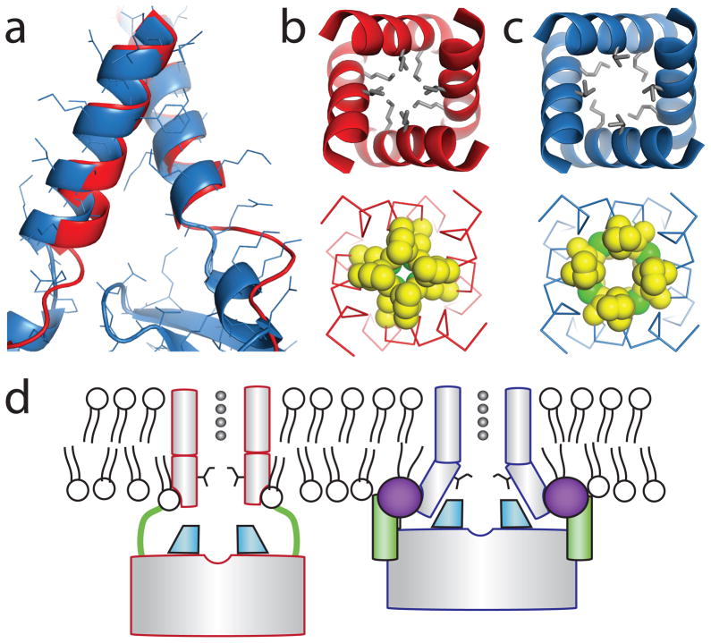
The regulation of ion channel activity by specific lipid molecules is widely recognized as an integral component of electrical signalling in cells. In particular, phosphatidylinositol 4,5-bisphosphate (PIP(2)), a minor yet dynamic phospholipid component of cell membranes, is known to regulate many different ion channels. PIP(2) is the primary agonist for classical inward rectifier (Kir2) channels, through which this lipid can regulate a cell's resting membrane potential. However, the molecular mechanism by which PIP(2) exerts its action is unknown. Here we present the X-ray crystal structure of a Kir2.2 channel in complex with a short-chain (dioctanoyl) derivative of PIP(2). We found that PIP(2) binds at an interface between the transmembrane domain (TMD) and the cytoplasmic domain (CTD). The PIP(2)-binding site consists of a conserved non-specific phospholipid-binding region in the TMD and a specific phosphatidylinositol-binding region in the CTD. On PIP(2) binding, a flexible expansion linker contracts to a compact helical structure, the CTD translates 6 Å and becomes tethered to the TMD and the inner helix gate begins to open. In contrast, the small anionic lipid dioctanoyl glycerol pyrophosphatidic acid (PPA) also binds to the non-specific TMD region, but not to the specific phosphatidylinositol region, and thus fails to engage the CTD or open the channel. Our results show how PIP(2) can control the resting membrane potential through a specific ion-channel-receptor-ligand interaction that brings about a large conformational change, analogous to neurotransmitter activation of ion channels at synapses.


Conflict of interest statement

The authors declare no competing financial interest.

Figures

Figure 1
Figure 1. Effect of a short chain PIP2 on Kir2.2
a, Endogenous PIP2 depletion causes “run down” of Kir2.2 channels in an excised inside out patch from Xenopus oocytes as shown by the three macroscopic current traces recorded with a voltage ramp from−80 to +80 mV immediately (green), 30 min (blue), and 50 min (black) after patch excision. b, The short chain PIP2 added to the bath solution (solid line with concentration indicated below) 30 min after patch excision partially rescued Kir2.2 channel activity. The bath was then perfused (dashed line) at time 40 min with ~1 ml/min bath solution for 3 min. c, X-ray crystal structures of apo (left, pdb 3JYC) and PIP2 bound (right, pdb 3SPI) Kir 2.2 tetramer (grey α-carbon traces) viewed from the side with the extracellular solution above. The lipid bilayer boundaries are shown as grey bars. Four PIP2 molecules are shown as sticks and colored according to atom type: carbon, yellow; phosphorous, orange; and oxygen, red. One PIP2 molecule in the same orientation as in figure 2a is outlined by a black box. Upon PIP2 binding the flexible linker between CTD and TMD consisting of two strands (highlighted green for one subunit, dotted line indicating disordered region in the crystal structure) form helical structures, and the CTD translates towards the TMD by 6 Å. A set of reference atoms (Asp72 and Lys220 α-carbons) are highlighted as blue spheres in each structure.
Figure 2
Figure 2. PIP2 binding site
a, A detailed view of the PIP2 binding site is shown in the same orientation as outlined in figure 1c. Helices (shown as ribbon) from different subunits are colored orange and cyan interior. Residues hydrogen bonded (dashed lines) to PIP2 are colored green, and residues stabilizing the PIP2 binding site in the CTD while lacking direct contact are colored blue. All side chains are shown as sticks. PIP2 is shown as sticks and colored according to atom type: carbon, yellow; phosphorous, orange; and oxygen, red. b, An amino acid sequence alignment of selected eukaryotic Kirs showing residues predicted from literature (blue outline) and not predicted (purple outline) to interact with PIP29,28. Residues with direct bonding interactions to PIP2 and with a structural role are highlighted with green and blue shade, respectively. The two residues serving as the inner helix gate are also highlighted with grey shade.
Figure 3
Figure 3. Conserved non-specific lipid binding site in Kirs
a, A grey α-carbon representation of Kir2.2 tetramer in complex with PPA, a small anionic lipid lacking an inositol ring. PPA bound Kir2.2 assumes a closed conformation similar to apo (pdb 3JYC) with the flexible linker elongated and the CTD unengaged. The four PPA molecules are shown as sticks and colored according to atom type: carbon, yellow; phosphorous, orange; and oxygen, red. b, A close-up view of the PPA binding site. PPA contacts Kir at the cytoplasmic end of the outer helix making strong interactions with the guanidiniums of R78 and R80 and the backbone amide nitrogens of the helix turn; similar to the interactions of the 1′ phosphate of PIP2. However, residues (blue sticks) for interacting with the PIP2 inositol 4′,5′ phosphate remain distant to the lipid binding site; R186 orients with its side chain pointing towards the ion conduction pathway. c, Superposition of PPA (colored the same as in panel a) and PIP2 (grey). The position of the 1′ phosphate in PIP2 is between the pyrophosphate of PPA.
Figure 4
Figure 4. A proposed mechanism of Kir2.2 activation by PIP2
a, Superposition of the TMD inner helices of the PIP2 bound (blue ribbon) and apo (red ribbon ) Kir2.2 structures. PIP2 binding results in a splaying of the helices near the helix bundle activation gate. b and c, comparison of the inner helix bundle gate in PPA bound Kir2.2 (b) and PIP2 bound Kir2.2 (c) viewed from the extracellular side. Side chains of the residues in the bundle crossing are represented as either grey sticks or space filling CPK models (carbon, yellow; and sulfur, green). d, A proposed mechanism for Kir2.2 activation by PIP2. PIP2(purple sphere) binds at an interface between the TMD (grey cylinder) and the CTD (grey rectangle) and induces a large conformational change: a flexible linker (green line) contracts to a compact helical structure (green cylinder), the CTD translates towards and becomes tethered to the TMD, the G-loop (cyan wedge) inserts into the TMD, and the inner helix activation gate opens.

References

    1. Dart C. Lipid microdomains and the regulation of ion channel function. J Physiol. 588:3169–3178. - PMC - PubMed
    1. Hilgemann DW, Feng S, Nasuhoglu C. The complex and intriguing lives of PIP2 with ion channels and transporters. Sci STKE. 2001;2001:re19. - PubMed
    1. Suh BC, Hille B. Regulation of ion channels by phosphatidylinositol 4,5-bisphosphate. Curr Opin Neurobiol. 2005;15:370–378. - PubMed
    1. Fujiwara Y, Kubo Y. Regulation of the desensitization and ion selectivity of ATP-gated P2X2 channels by phosphoinositides. J Physiol. 2006;576:135–149. - PMC - PubMed
    1. Logothetis DE, Jin T, Lupyan D, Rosenhouse-Dantsker A. Phosphoinositide-mediated gating of inwardly rectifying K(+) channels. Pflügers Archiv : European journal of physiology. 2007;455:83–95. - PubMed

Publication types

MeSH terms

Substances