. 2012 Mar 9;287(11):7915-24.
doi: 10.1074/jbc.M111.305078. Epub 2012 Jan 18.

Functional and structural characterization of α-(1->2) branching sucrase derived from DSR-E glucansucrase

Affiliations

Functional and structural characterization of α-(1->2) branching sucrase derived from DSR-E glucansucrase

Yoann Brison et al. J Biol Chem. .

Abstract

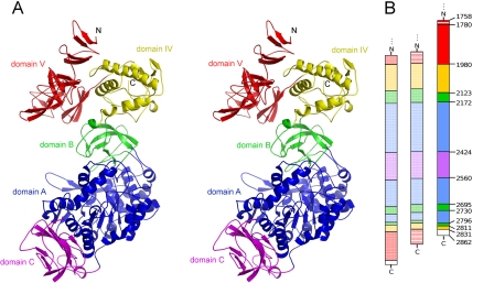
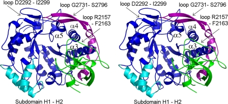
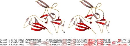
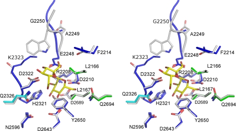
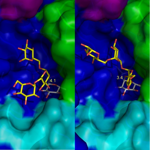
ΔN(123)-glucan-binding domain-catalytic domain 2 (ΔN(123)-GBD-CD2) is a truncated form of the bifunctional glucansucrase DSR-E from Leuconostoc mesenteroides NRRL B-1299. It was constructed by rational truncation of GBD-CD2, which harbors the second catalytic domain of DSR-E. Like GBD-CD2, this variant displays α-(1→2) branching activity when incubated with sucrose as glucosyl donor and (oligo-)dextran as acceptor, transferring glucosyl residues to the acceptor via a ping-pong bi-bi mechanism. This allows the formation of prebiotic molecules containing controlled amounts of α-(1→2) linkages. The crystal structure of the apo α-(1→2) branching sucrase ΔN(123)-GBD-CD2 was solved at 1.90 Å resolution. The protein adopts the unusual U-shape fold organized in five distinct domains, also found in GTF180-ΔN and GTF-SI glucansucrases of glycoside hydrolase family 70. Residues forming subsite -1, involved in binding the glucosyl residue of sucrose and catalysis, are strictly conserved in both GTF180-ΔN and ΔN(123)-GBD-CD2. Subsite +1 analysis revealed three residues (Ala-2249, Gly-2250, and Phe-2214) that are specific to ΔN(123)-GBD-CD2. Mutation of these residues to the corresponding residues found in GTF180-ΔN showed that Ala-2249 and Gly-2250 are not directly involved in substrate binding and regiospecificity. In contrast, mutant F2214N had lost its ability to branch dextran, although it was still active on sucrose alone. Furthermore, three loops belonging to domains A and B at the upper part of the catalytic gorge are also specific to ΔN(123)-GBD-CD2. These distinguishing features are also proposed to be involved in the correct positioning of dextran acceptor molecules allowing the formation of α-(1→2) branches.


Figures

FIGURE 1.
FIGURE 1.
A, anomeric region of 1H NMR spectra obtained at 298 K for purified α-(1→2) branched dextrans. Black, dextran 70-kDa standard; blue, dextran α-(1→2) branched at 11%; pink, dextran α-(1→2) branched at 19%; orange, dextran α-(1→2) branched at 33%; green, dextrans α-(1→2) branched between 35 and 37%. B, percentage of α-(1→2) linkage as a function of [sucrose]/[dextran] ([Suc]/[Dex]) molar ratios used for the acceptor reactions. Empty and filled circles correspond to values obtained after 1H NMR and HPLC measurement, respectively, of the α-(1→2) linkage content in dextrans synthesized by ΔN123-GBD-CD2. Red crosses, 1H NMR results for dextrans branched by GBD-CD2 (12). C, effects of the [sucrose]/[dextran] molar ratio on α-(1→2) branched dextran yields. The reactions were carried out at 292 mm sucrose and various dextran concentrations. Main final reaction products are residual sucrose, glucose (from sucrose hydrolysis), leucrose (from fructose glucosylation), and α-(1→2) branched dextran.
FIGURE 2.
FIGURE 2.
A, stereo view of ΔN123-GBD-CD2 domain organization. Magenta, domain C; blue, domain A, which includes the (β/α)8 barrel; green, domain B; yellow, domain IV; red, domain V. B, schematic representation of the domain arrangement along the polypeptide chains of crystallized GH70 glucansucrases, from left to right, GTF180-ΔN, GTF-SI, and ΔN123-GBD-CD2. The color code is identical to that for A. Striped red, parts of domains V of GTF-SI and ΔN123-GBD-CD2, which are not visible in the electron density map; white, purification tag.
FIGURE 3.
FIGURE 3.
Stereo view of the secondary structure elements of ΔN123-GBD-CD2 domains A and B. For domain A, blue shows the (β/α)8 barrel, cyan shows the subdomain H1-H2, and purple shows the loop Gly-2731 to Ser-2796 protruding from domain B and contributing to domain A. Domain B is green.
FIGURE 4.
FIGURE 4.
Upper panel, stereo view of the secondary structure elements of domain V (truncated glucan-binding domain) of ΔN123-GBD-CD2. The three-stranded β-sheets and β-hairpins of the three subdomains are represented in red and salmon, respectively. Lower panel, sequence alignment of the domain V. The underlined and colored residues are β-strands; N-terminal residues in italics are not visible in the electron density map.
FIGURE 5.
FIGURE 5.
Stereo view of subsites −1 and +1 of ΔN123-GBD-CD2, with sucrose from the GTF180-ΔN-sucrose complex superimposed. The catalytic residues are Asp-2210 (nucleophile), Glu-2248 (acid/base), and Asp-2322 (transition state stabilizer). Sucrose is shown with yellow carbons. Residues of the inactive GTF180-ΔN mutant (D1025N) that interact with sucrose (8) are represented in gray. The carbon atoms of their structural equivalents in ΔN123-GBD-CD2 are shown in blue (domain A), cyan (subdomain H1-H2), and green (domain B).
FIGURE 6.
FIGURE 6.
Isomaltotriose docking in the catalytic groove of the modeled glucosyl-enzyme intermediate of ΔN123-GBD-CD2. Two binding modes were found that allow glucosylation through α-(1→2) linkage formation onto the central glucosyl unit (A) or the nonreducing end extremity (B). The covalently linked glucosyl unit in subsite −1 is represented in gray, and isomaltotriose is in yellow. The distances between O2 atom of isomaltotriose and C1 atom of the glucosyl enzyme intermediate (in Å) are shown in yellow. Domain coloring is as in Fig. 3.
FIGURE 7.
FIGURE 7.
Stereo view of the catalytic gorges of GTF180-ΔN and ΔN123-GBD-CD2. For clarity, only different residues or residues adopting different conformations are shown. Backbone atoms of ΔN123-GBD-CD2 are depicted in light blue. Residues from domain A of ΔN123-GBD-CD2 are depicted in blue, cyan, and purple (see Fig. 3). Residues from domain B of ΔN123-GBD-CD2 are shown in green. Gray residues belong to GTF180-ΔN. Sucrose from GTF180-ΔN-sucrose complex (Protein Data Bank entry 3HZ3) in subsites −1 and +1 is represented as yellow carbons.

References

    1. Henrissat B., Bairoch A. (1996) Updating the sequence-based classification of glycosyl hydrolases. Biochem. J. 316, 695–696 - PMC - PubMed
    1. Monsan P., Remaud-Siméon M., André I. (2010) Transglucosidases as efficient tools for oligosaccharide and glucoconjugate synthesis. Curr. Opin. Microbiol. 13, 293–300 - PubMed
    1. MacGregor E. A., Janecek S., Svensson B. (2001) Relationship of sequence and structure to specificity in the α-amylase family of enzymes. Biochim. Biophys. Acta 1546, 1–20 - PubMed
    1. Moulis C., Joucla G., Harrison D., Fabre E., Potocki-Veronese G., Monsan P., Remaud-Simeon M. (2006) Understanding the polymerization mechanism of glycoside-hydrolase family 70 glucansucrases. J. Biol. Chem. 281, 31254–31267 - PubMed
    1. MacGregor E. A., Jespersen H. M., Svensson B. (1996) A circularly permuted α-amylase-type α/β-barrel structure in glucan-synthesizing glucosyltransferases. FEBS Lett. 378, 263–266 - PubMed

Publication types

LinkOut - more resources