. 2011 Dec 2;334(6060):1289-93.
doi: 10.1126/science.1213782. Epub 2011 Oct 27.

Increasing the potency and breadth of an HIV antibody by using structure-based rational design

Affiliations

Increasing the potency and breadth of an HIV antibody by using structure-based rational design

Ron Diskin et al. Science. .

Abstract

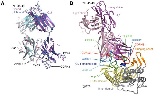
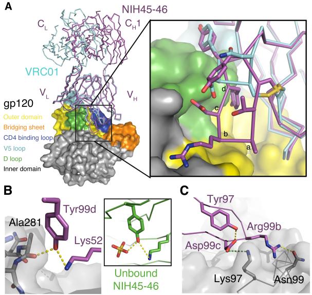
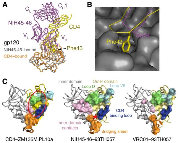
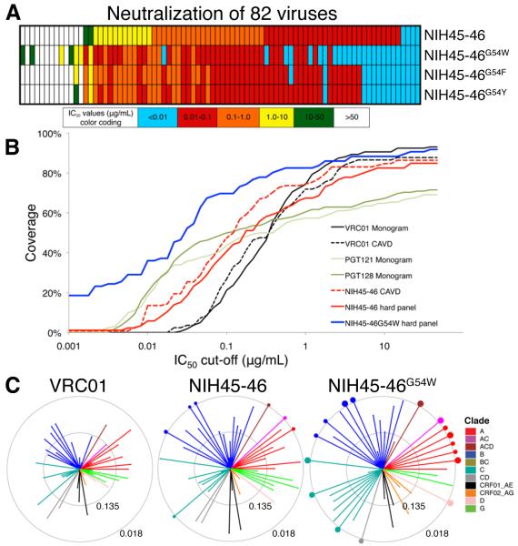
Antibodies against the CD4 binding site (CD4bs) on the HIV-1 spike protein gp120 can show exceptional potency and breadth. We determined structures of NIH45-46, a more potent clonal variant of VRC01, alone and bound to gp120. Comparisons with VRC01-gp120 revealed that a four-residue insertion in heavy chain complementarity-determining region 3 (CDRH3) contributed to increased interaction between NIH45-46 and the gp120 inner domain, which correlated with enhanced neutralization. We used structure-based design to create NIH45-46(G54W), a single substitution in CDRH2 that increases contact with the gp120 bridging sheet and improves breadth and potency, critical properties for potential clinical use, by an order of magnitude. Together with the NIH45-46-gp120 structure, these results indicate that gp120 inner domain and bridging sheet residues should be included in immunogens to elicit CD4bs antibodies.


Figures

Fig. 1
Fig. 1
Crystal structures of NIH45-46 Fab alone and bound to gp120. (A) Superimposition of the structures of the free (blue heavy chain and cyan light chain) and bound (magenta heavy chain and pink light chain) NIH45-46 Fab. RMSDs for free and bound VH-VH and VL-VL superimpositions (123 and 99 Cα atoms, respectively) are each 0.5 Å. The location of an extra disulfide bond that joins Cys32–Cys98 in VH is marked with an asterisk and N-linked carbohydrate attached to Asn70 of VL is shown as sticks. Arrows point to slightly different conformations in CDRL1 and CDRH3 in bound and free NIH45-46. Highlighted sidechains, Tyr89CDRL3 and Tyr74FWRH3, adopt notably different conformations upon binding gp120. (B) Structure of the NIH45-46–gp120 complex. Ribbon diagram of the NIH45-46 Fab (magenta and pink for the heavy and light chains, respectively) complexed with the inner (yellow) and outer (gray) domains of the 93TH053 gp120 core, which lacks three variable loops (V1-V2 and V3) and has N- and C-terminal truncations (25). Lines point to structural features of the gp120 core discussed in the text.
Fig. 2
Fig. 2
Interactions of the NIH45-46 insertion with gp120. (A) Superimposition of the gp120 portions of VRC01–93TH053 (PDB 3NGB) and NIH45-46–93TH053 structures. The Fabs are shown as magenta (NIH45-46) or cyan (VRC01) wire, and gp120 is shown as a surface with the color scheme used in Fig. 1B. The region surrounding the four-residue insertion in the NIH45-46 CDRH3 (residues 99a – 99d) is boxed. Inset: Close-up of the boxed region in which the NIH45-46 insertion residues are labeled alphabetically to correspond with residues 99a – 99d. The sidechains of relevant CDRH3 residues are shown as sticks. (B) Hydrogen bond network between the mainchain carbonyl oxygen of Ala281gp120, Tyr99dNIH45-46 in CDRH3, and Lys52NIH45-46 in CDRH2. Yellow dots represent hydrogen bonds. The conformation of Tyr99dNIH45-46 is also stabilized by a hydrogen bond with Lys52NIH45-46 in unbound NIH45-46 (inset) in which a sulfate ion (yellow) from the crystallization solution substitutes for the interaction with Ala281gp120. (C) Electrostatic interaction between Asp99cNIH45-46 and Lys97gp120 (green dots) is shown with Asp99cNIH45-46-Tyr97NIH45-46 and Arg99bNIH45-46–Asn99gp120 hydrogen bonds (yellow dots).
Fig. 3
Fig. 3
NIH45-46 mimicry of CD4 binding. (A) Superimposition of NIH45-46–gp120 (magenta and gray, respectively) and CD4–gp120 (yellow and orange, respectively) (31) calculated based on gp120 Cα positions. Phe43CD4 is shown using spheres. (B) Close-up of the CDRH2 loop of NIH45-46 (magenta) and CDR2-like loop of CD4 (yellow) interacting with gp120 (gray surface). Phe43CD4 (labeled) inserts into a hydrophobic pocket on gp120. The closest corresponding residue on NIH45-46 (Gly54NIH45-46) is labeled. (C) Comparison of contacts on gp120 made by CD4, NIH45-46, and VRC01 (left, middle and right respectively). Residues at each contact interface are highlighted on the gp120 structure (colored as in Fig. 1B) as a surface enclosing the contact residues. The location of the Phe43 cavity targeted by the tryptophan substitution in NIH45-46G54W is indicated on the middle panel with a red asterisk. The approximate location of the gp120 region defined as the initial site of CD4 attachment (25, 26) is indicated with a red oval in the left panel.
Fig. 4
Fig. 4
Increased neutralization potency of NIH45-46G54W. (A) Schematic comparing neutralization potencies for NIH45-46 and three position 54 mutants, including NIH45-46G54W. IC50 values are color-coded as shown. Numerical values for the same 82 strains are shown in Table S5 (IC50 values) and Table S6 (IC80 values). (B) Graphical comparisons of neutralization coverage and potency. The y-axis shows the cumulative frequency of IC50 values up to the concentration shown on the x-axis. The curves for VRC01, the only antibody tested in both the CAVD panel (a panel of 118 viral strains (16)) and the Monogram panel (a panel of 162 viral strains (18)), are similar, although the CAVD panel includes a few more resistant strains (compare the solid and dotted black lines). NIH45-46 (red dotted line) was tested on the CAVD panel and is more potent than VRC01 and broader than the PGT Abs. NIH45-46G54W (solid blue line) was tested with NIH45-46 (solid red line) on a panel of 82 viral strains chosen to include many NIH45-46 resistant and poorly neutralized strains (hard panel; Tables S5,S6), thus resulting in lower coverage and lower potency for NIH45-46 when evaluated using the hard panel versus using the CAVD panel (compare the solid and dotted red lines). The improved potency and breadth of NIH45-46G54W versus NIH45-46 against the hard panel are apparent. Even when evaluated using a difficult to neutralize panel of viruses, NIH45-46G54W is the most potent and broadly neutralizing antibody described to date. (C) Neutralization summary spider graphs comparing IC50 values for VRC01, NIH45-46, and NIH45-46G54W for 65 common viruses that were tested in this work or previously (16). Each color represents a different HIV clade. Length of lines and size of circles are inversely proportional to IC50. The distance between the outer and inner circle and the distance from the inner circle to the center of a spider graph each span two natural logs in IC50 concentration (concentrations in μg/mL are indicated on each circle). Dots on the outer circle indicate strains with IC50 values less than 0.018 μg/mL whose lines were truncated in the graph. The size of each dot is inversely proportional to the IC50 value.

References

    1. Fauci AS, et al. Science. 2008 Jul 25;321:530. - PubMed
    1. McElrath MJ, Haynes BF. Immunity. 2010 Oct 29;33:542. - PMC - PubMed
    1. Karlsson Hedestam GB, et al. Nat Rev Microbiol. 2008 Feb;6:143. - PubMed
    1. McMichael AJ, Borrow P, Tomaras GD, Goonetilleke N, Haynes BF. Nat Rev Immunol. 2010 Jan;10:11. - PMC - PubMed
    1. Zolla Pazner S, Cardozo T. Nature reviews. Immunology. 2010;10:527. - PMC - PubMed

Publication types

MeSH terms