Comparative Study
. 2011 Dec 11;481(7380):219-23.
doi: 10.1038/nature10691.

An unanticipated architecture of the 750-kDa α6β6 holoenzyme of 3-methylcrotonyl-CoA carboxylase

Affiliations
Comparative Study

An unanticipated architecture of the 750-kDa α6β6 holoenzyme of 3-methylcrotonyl-CoA carboxylase

Christine S Huang et al. Nature. .

Abstract

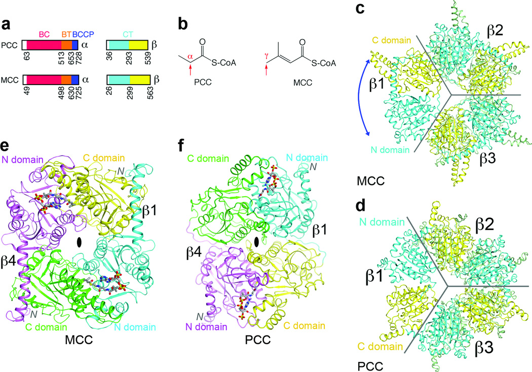
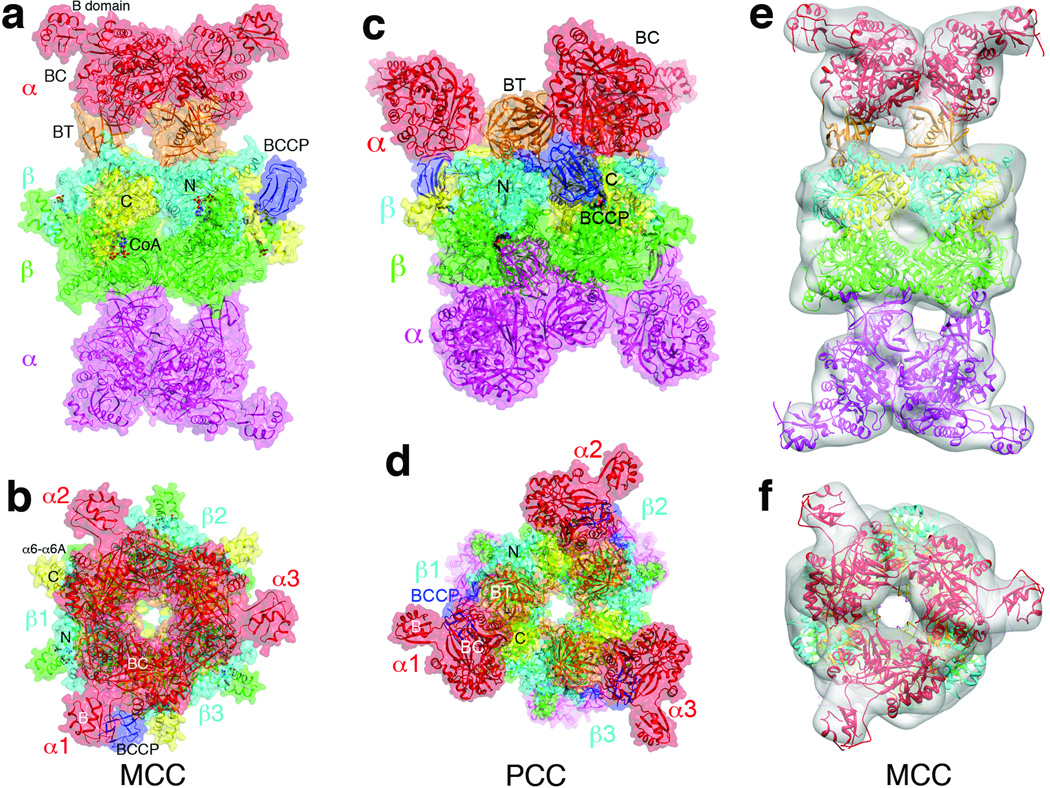
3-Methylcrotonyl-CoA carboxylase (MCC), a member of the biotin-dependent carboxylase superfamily, is essential for the metabolism of leucine, and deficient mutations in this enzyme are linked to methylcrotonylglycinuria (MCG) and other serious diseases in humans. MCC has strong sequence conservation with propionyl-CoA carboxylase (PCC), and their holoenzymes are both 750-kilodalton (kDa) α(6)β(6) dodecamers. Therefore the architecture of the MCC holoenzyme is expected to be highly similar to that of PCC. Here we report the crystal structures of the Pseudomonas aeruginosa MCC (PaMCC) holoenzyme, alone and in complex with coenzyme A. Surprisingly, the structures show that the architecture and overall shape of PaMCC are markedly different when compared to PCC. The α-subunits show trimeric association in the PaMCC holoenzyme, whereas they have no contacts with each other in PCC. Moreover, the positions of the two domains in the β-subunit of PaMCC are swapped relative to those in PCC. This structural information establishes a foundation for understanding the disease-causing mutations of MCC and provides new insights into the catalytic mechanism and evolution of biotin-dependent carboxylases. The large structural differences between MCC and PCC also have general implications for the relationship between sequence conservation and structural similarity.


Figures

Figure 1
Figure 1. The domains of MCCβ are swapped compared to PCCβ
(a). Domain organization of human MCC and PCC. (b). Distinct carboxylation targets of MCC and PCC, indicated by the red arrow. (c). Crystal structure of the β6 hexamer of PaMCC. The subunit beneath β1 is omitted for clarity, and the other two subunits in the bottom layer are colored in green. The blue arrow indicates the swapping of the positions of the N and C domains relative to PCCβ. Gray lines mark the boundaries of the subunits. (d). Structure of Roseobacter denitrificans PCCβ . (e). Structure of the β2 dimer of PaMCC. The N and C domains of the subunit in the bottom layer (β4) are colored in magenta and green, respectively. (f). Structure of the β2 dimer of PCC. All the structure figures were produced with PyMOL (www.pymol.org) unless stated otherwise.
Figure 2
Figure 2. The MCC holoenzyme has a strikingly different architecture compared to PCC
(a). Structure of the CoA complex of PaMCC holoenzyme, side view. Domains in the α and β subunits in the top half of the structure are colored as in Fig. 1a. The α and β subunits in the bottom half are colored in magenta and green, respectively. The molecular surface is shown in a semi-transparent rendering. (b). Structure of the PaMCC holoenzyme, top view. (c). Structure of the PCC holoenzyme , side view. (d). Structure of the PCC holoenzyme, top view. (e). EM reconstruction of PaMCC at 12 Å resolution, side view. The crystal structure of the PaMCC free enzyme can be readily fit into the EM density. (f). EM reconstruction of PaMCC, top view. Panels e and f were produced with Chimera .
Figure 3
Figure 3. The BT domain mediates interactions in the MCC holoenzyme
(a). Overlay of the structure of PaMCC BT domain (in orange) with that of PCC (gray). A large conformational difference for the hook is visible. The exact positions of many of the β-strands are different as well. (b). The BT domain (orange) contacts a β subunit (β1, N domain in cyan, C domain yellow) as well as a neighboring α subunit (α2, red) in the PaMCC holoenzyme. (c). Detailed interactions between the hook of the BT domain and the β subunit in PaMCC. Three disease-causing mutation sites near this interface are labeled in red. For stereo version of the panels, please see Supplementary Figs. 9, 10.
Figure 4
Figure 4. Molecular basis for catalysis and disease-causing mutations in the MCC holoenzyme
(a). The BC and CT active sites (indicated with the asterisks) are separated by 80 Å in the PaMCC holoenzyme. Molecular surfaces of one α subunit and two β2 dimers are shown. The position of ADP bound to the BC subunit of E. coli acetyl-CoA carboxylase indicates the BC active site. Helices α6-α6A are shown as a ribbon in order to make CoA visible. (b). Schematic drawing of the active site of the β subunit. Biotin (black) and the modeled conformation of methylcrotonyl-CoA (gray) are shown as stick models. Residue Ala218 is the site of a disease-causing mutation. For stereo version of this panel, please see Supplementary Fig. 14. (c). Disease-causing mutation sites are shown as spheres in the PaMCC structure. The mutations are distributed throughout the holoenzyme.

References

    1. Baumgartner MR, et al. The molecular basis of human 3-methylcrotonyl-CoA carboxylase deficiency. J. Clin. Investig. 2001;107:495–504. - PMC - PubMed
    1. Gallardo ME, et al. The molecular basis of 3-methylcrotonylglycinuria, a disorder of leucine metabolism. Am. J. Hum. Genet. 2001;68:334–346. - PMC - PubMed
    1. Holzinger A, et al. Cloning of the human MCCA and MCCB genes and mutations therein reveal the molecular cause of 3-methylcrotonyl-CoA carboxylase deficiency. Human Mol. Gen. 2001;10:1299–1306. - PubMed
    1. Desviat LR, et al. Functional analysis of MCCA and MCCB mutations causing methylcrotonylglycinuria. Mol. Gen. Metab. 2003;80:315–320. - PubMed
    1. Wakil SJ, Stoops JK, Joshi VC. Fatty acid synthesis and its regulation. Ann. Rev. Biochem. 1983;52:537–579. - PubMed

Publication types

MeSH terms