. 2016 Nov 11;291(46):24065-24075.
doi: 10.1074/jbc.M116.756965. Epub 2016 Sep 27.

Structures of Proline Utilization A (PutA) Reveal the Fold and Functions of the Aldehyde Dehydrogenase Superfamily Domain of Unknown Function

Affiliations

Structures of Proline Utilization A (PutA) Reveal the Fold and Functions of the Aldehyde Dehydrogenase Superfamily Domain of Unknown Function

Min Luo et al. J Biol Chem. .

Abstract

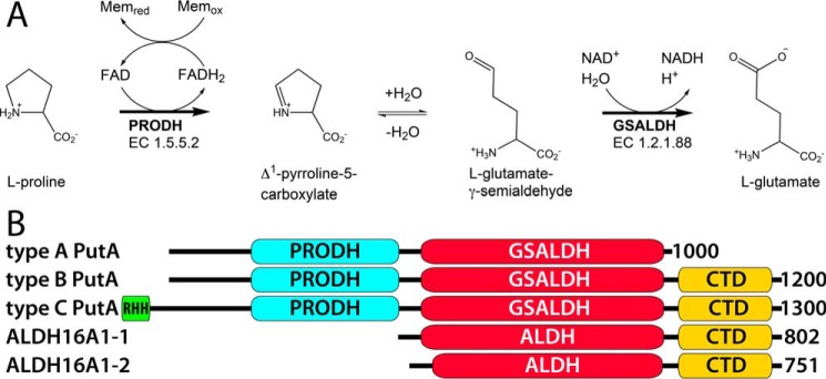
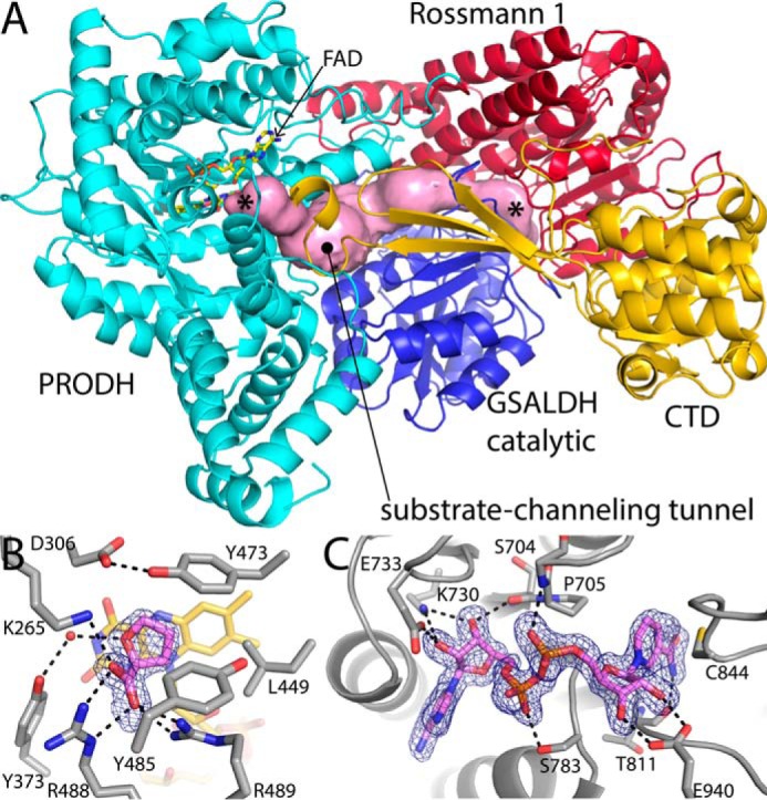
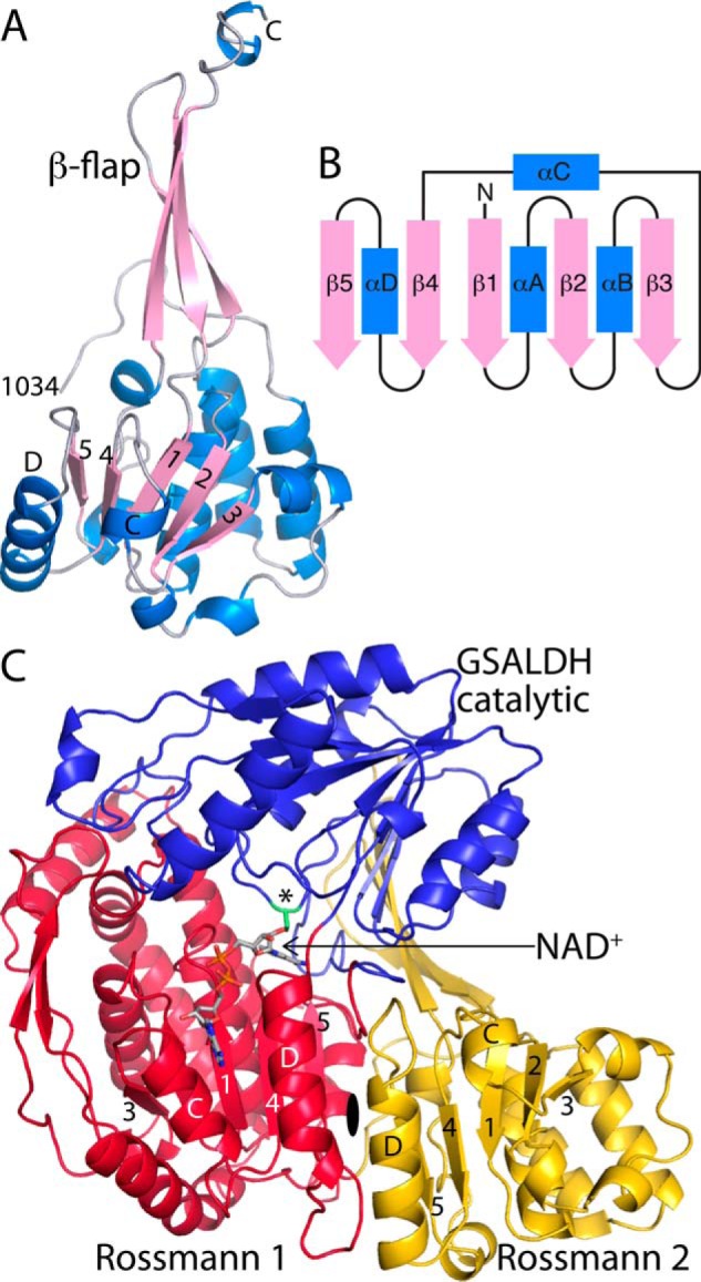
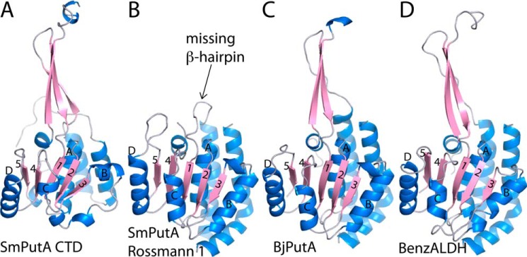
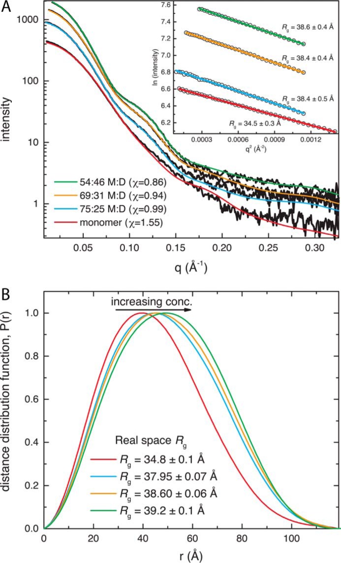
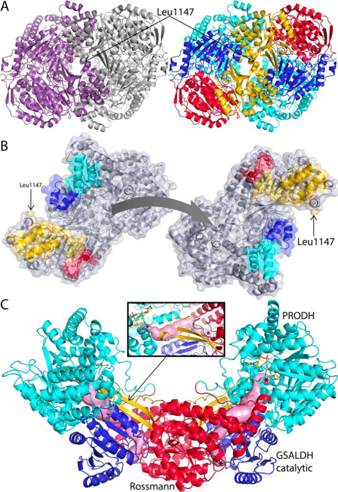
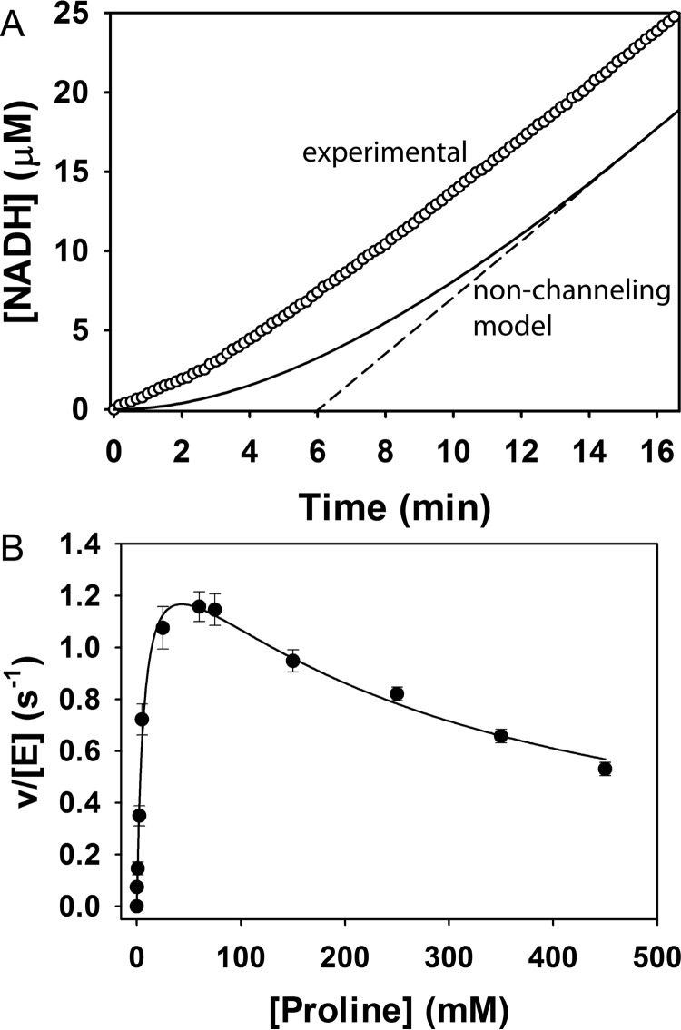
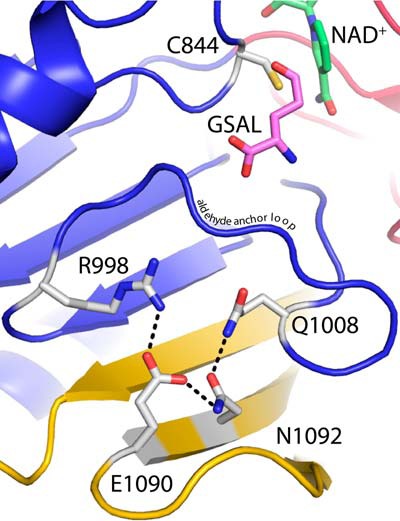
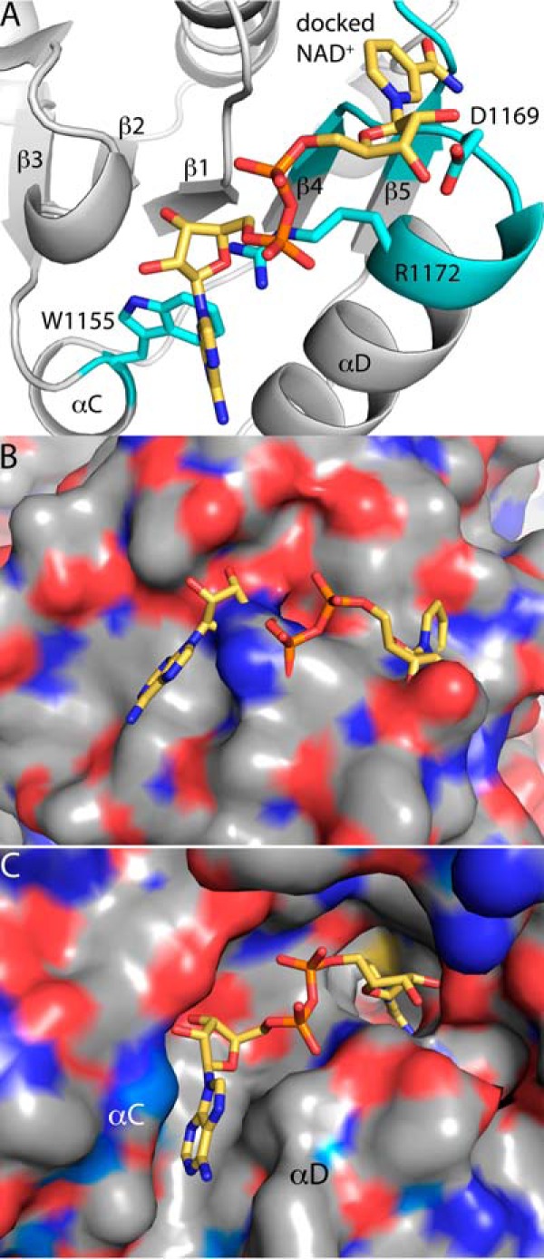
Aldehyde dehydrogenases (ALDHs) catalyze the NAD(P)+-dependent oxidation of aldehydes to carboxylic acids and are important for metabolism and detoxification. Although the ALDH superfamily fold is well established, some ALDHs contain an uncharacterized domain of unknown function (DUF) near the C terminus of the polypeptide chain. Herein, we report the first structure of a protein containing the ALDH superfamily DUF. Proline utilization A from Sinorhizobium meliloti (SmPutA) is a 1233-residue bifunctional enzyme that contains the DUF in addition to proline dehydrogenase and l-glutamate-γ-semialdehyde dehydrogenase catalytic modules. Structures of SmPutA with a proline analog bound to the proline dehydrogenase site and NAD+ bound to the ALDH site were determined in two space groups at 1.7-1.9 Å resolution. The DUF consists of a Rossmann dinucleotide-binding fold fused to a three-stranded β-flap. The Rossmann domain resembles the classic ALDH superfamily NAD+-binding domain, whereas the flap is strikingly similar to the ALDH superfamily dimerization domain. Paradoxically, neither structural element performs its implied function. Electron density maps show that NAD+ does not bind to the DUF Rossmann fold, and small-angle X-ray scattering reveals a novel dimer that has never been seen in the ALDH superfamily. The structure suggests that the DUF is an adapter domain that stabilizes the aldehyde substrate binding loop and seals the substrate-channeling tunnel via tertiary structural interactions that mimic the quaternary structural interactions found in non-DUF PutAs. Kinetic data for SmPutA indicate a substrate-channeling mechanism, in agreement with previous studies of other PutAs.

Keywords: X-ray crystallography; aldehyde dehydrogenase superfamily; enzyme kinetics; flavoprotein; nicotinamide adenine dinucleotide (NAD); oligomerization; proline catabolism; proline utilization A; protein domain; small-angle X-ray scattering (SAXS).


Figures

FIGURE 1.
FIGURE 1.
The reactions of proline catabolism and the domains of PutA and ALDH16A1. A, the reactions catalyzed by PutA. B, domain diagrams of PutAs and ALDH16A1. The small N-terminal domain in type C PutAs is a ribbon-helix-helix DNA-binding domain.
FIGURE 2.
FIGURE 2.
Structure of SmPutA. A, cartoon drawing of the protomer. The PRODH module is colored cyan. The Rossmann NAD+-binding and catalytic domains of the GSALDH module are colored red and blue, respectively. The CTD is colored gold. The pink surface represents the substrate-channeling tunnel. The asterisks indicate the locations of the two active sites in the tunnel, with the PRODH site on the left and the GSALDH site on the right. B, electron density and interactions for the proline analog THFA bound to the SmPutA PRODH active site (space group P21, chain A). The cage represents a simulated annealing σA-weighted FoFc omit map (3.0 σ). C, electron density and interactions for the NAD+ bound to SmPutA (space group P21). The cage represents a simulated annealing σA-weighted FoFc omit map (3.0 σ).
FIGURE 3.
FIGURE 3.
The fold and tertiary structural interactions of the CTD. A, cartoon drawing of the CTD. Selected β-strands and α-helices of the Rossmann domain are indicated. B, topology diagram of the Rossmann dinucleotide-binding fold found in ALDHs. C, tertiary structural interactions between the CTD (gold) and the GSALDH module. The asterisk indicates catalytic Cys844.
FIGURE 4.
FIGURE 4.
Comparison of the CTD to ALDH superfamily domains. A, the CTD of SmPutA. B, the Rossmann NAD+-binding domain in the GSALDH module of SmPutA. C, the Rossmann NAD+-binding and oligomerization domains of BjPutA (PDB code 3HAZ). D, the Rossmann and oligomerization domains of benzaldehyde dehydrogenase (PDB code 3R64). In all panels, the diagnostic strands and helices of the Rossmann fold are indicated.
FIGURE 5.
FIGURE 5.
SAXS analysis of SmPutA. A, SAXS curves from samples at different protein concentrations. The black curves represent the experimental data. The smooth curves represent theoretical SAXS curves calculated from atomic models. The inset shows Guinier plots. B, distance distribution functions calculated from the SAXS curves.
FIGURE 6.
FIGURE 6.
A novel ALDH dimer. A, cartoon representation of the SmPutA dimer viewed down the 2-fold axis. On the left, the two protomers have different colors. On the right, the protein is color-coded by modules/domains as in Fig. 2A: PRODH, cyan; Rossmann 1, red; GSALDH catalytic, blue; and CTD, gold. B, the separated protomers of the SmPutA dimer. The interaction surfaces are color-coded according to modules/domains: PRODH, cyan; Rossmann 1, red; GSALDH catalytic, blue; and CTD, gold. C, the traditional ALDH mode of dimerization as seen in BjPutA (a type A PutA, PDB code 3HAZ). The domains are colored as follows: PRODH, cyan; Rossmann NAD+-binding domain, red; GSALDH catalytic, blue; and oligomerization flap, gold. The pink surface represents the substrate-channeling tunnels. The inset shows a close-up view of the oligomerization flap of one protomer covering the substrate-channeling tunnel of the opposite protomer. The asterisks indicate the locations of the two active sites. Note that the quaternary structural interactions in BjPutA resemble the tertiary structural interactions of the β-flap in SmPutA (Fig. 2A).
FIGURE 7.
FIGURE 7.
Kinetics of substrate channeling. A, transient time analysis of SmPutA. The circles show NADH production from SmPutA (0.25 μm) with 40 mm proline, 200 μm CoQ1, and 200 μm NAD+, pH 7.5. The solid curve shows the predicted NADH formation using a two-enzyme nonchanneling model of the SmPutA PRODH-GSALDH coupled reaction (Equation 1). Linear extrapolation of the nonchanneling model as shown by the dashed line yields a transient time of 6 min. B, initial velocity of the coupled PRODH-GSALDH reaction with varied proline concentration and fixed CoQ1 (300 μm) and NAD+ (200 μm). Non-linear least squares fit of the data to a substrate inhibition model as shown gave best fit parameters of kcat = 1.6 ± 0.1 s−1, Km = 7 ± 1 mm, and Ki = 263 ± 36 mm proline.
FIGURE 8.
FIGURE 8.
Proximity of the β-flap of the CTD to the GSALDH active site. The cartoon is color-coded according to domains: Rossmann 1, red; GSALDH catalytic, blue; and CTD, gold. A model of GSAL has been docked into SmPutA based on a structure of GSALDH complexed with glutamate (PDB code 3V9K).
FIGURE 9.
FIGURE 9.
Structural explanation for why the CTD does not bind NAD+. A, a model of NAD+ from the GSALDH domain has been docked to the CTD in the canonical position for Rossmann dinucleotide-binding domains. Residues predicted to clash NAD+ are in cyan. B, the surface of the CTD where NAD+ would be expected to bind. NAD+ is docked into the canonical position for Rossmann dinucleotide-binding domains. Note the absence of a well defined pocket for NAD+. C, the surface of the bona fide NAD+-binding site of the Rossmann 1 domain of SmPutA. Note the presence of a pocket that is complementary in shape to the active conformation of NAD+.

References

    1. Liu Z. J., Sun Y. J., Rose J., Chung Y. J., Hsiao C. D., Chang W. R., Kuo I., Perozich J., Lindahl R., Hempel J., and Wang B. C. (1997) The first structure of an aldehyde dehydrogenase reveals novel interactions between NAD and the Rossmann fold. Nat. Struct. Biol. 4, 317–326 - PubMed
    1. Tanner J. J. (2008) Structural biology of proline catabolism. Amino Acids 35, 719–730 - PMC - PubMed
    1. Tanner J. J., and Becker D. F. (2013) PutA and proline metabolism. In Handbook of Flavoproteins: Volume 1. Oxidases, Dehydrogenases and Related Systems (Hille R., Miller S. M., and Palfey B. A., eds) pp. 31–56, De Gruyter, Berlin
    1. Srivastava D., Schuermann J. P., White T. A., Krishnan N., Sanyal N., Hura G. L., Tan A., Henzl M. T., Becker D. F., and Tanner J. J. (2010) Crystal structure of the bifunctional proline utilization A flavoenzyme from Bradyrhizobium japonicum. Proc. Natl. Acad. Sci. U.S.A. 107, 2878–2883 - PMC - PubMed
    1. Singh H., Arentson B. W., Becker D. F., and Tanner J. J. (2014) Structures of the PutA peripheral membrane flavoenzyme reveal a dynamic substrate-channeling tunnel and the quinone-binding site. Proc. Natl. Acad. Sci. U.S.A. 111, 3389–3394 - PMC - PubMed

LinkOut - more resources