. 2014 Mar 18;111(11):4049-54.
doi: 10.1073/pnas.1321562111. Epub 2014 Mar 3.

Structural basis for gating mechanisms of a eukaryotic P-glycoprotein homolog

Affiliations

Structural basis for gating mechanisms of a eukaryotic P-glycoprotein homolog

Atsushi Kodan et al. Proc Natl Acad Sci U S A. .

Abstract

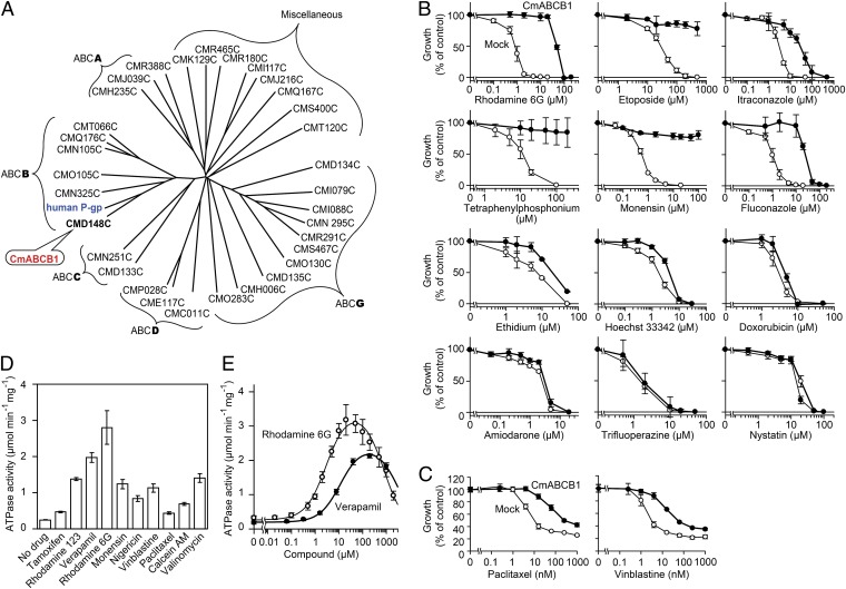
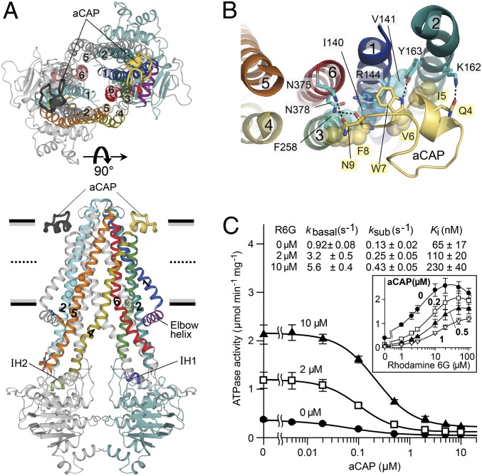
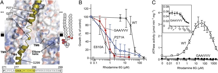
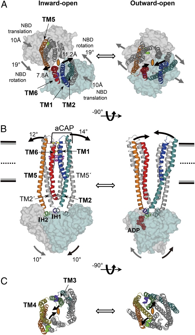
P-glycoprotein is an ATP-binding cassette multidrug transporter that actively transports chemically diverse substrates across the lipid bilayer. The precise molecular mechanism underlying transport is not fully understood. Here, we present crystal structures of a eukaryotic P-glycoprotein homolog, CmABCB1 from Cyanidioschyzon merolae, in two forms: unbound at 2.6-Å resolution and bound to a unique allosteric inhibitor at 2.4-Å resolution. The inhibitor clamps the transmembrane helices from the outside, fixing the CmABCB1 structure in an inward-open conformation similar to the unbound structure, confirming that an outward-opening motion is required for ATP hydrolysis cycle. These structures, along with site-directed mutagenesis and transporter activity measurements, reveal the detailed architecture of the transporter, including a gate that opens to extracellular side and two gates that open to intramembranous region and the cytosolic side. We propose that the motion of the nucleotide-binding domain drives those gating apparatuses via two short intracellular helices, IH1 and IH2, and two transmembrane helices, TM2 and TM5.

Keywords: ABC transporter; X-ray crystallography; macrocyclic peptide; membrane protein; multidrug resistance.


Conflict of interest statement

The authors declare no conflict of interest.

Figures

Fig. 1.
Fig. 1.
CmABCB1 is an ABC multidrug transporter. (A) Dendrogram of ABC transporters from C. merolae and human P-gp (ABCB1). Among 32 ABC proteins from C. merolae, CMD148C is the most similar to human P-gp and was therefore designated as CmABCB1. This dendrogram is based on multiple sequence alignments of the full amino acid sequences of ABC transporters. Alignments were performed using with ClustalW and visualized using Drawtree. (B) Drug-susceptibility assay in S. cerevisiae AD1-8u cells. AD1-8u cells expressing WT CmABCB1 (●) were grown in various concentrations of drugs. For each drug assayed, mock-transfected AD1-8u cells (○) were also grown as controls. Data are means ± SD (n = 5). (C) Drug-susceptibility assay in HEK293 cells. Mock-transfected HEK293 (○) and CmABCB1-expressing HEK293 cells (●) were incubated with paclitaxel or vinblastine, and viability was measured by the 3-(4,5-dimethylthiazol-2-yl)-2,5-diphenyl tetrazolium bromide (MTT) assay. Data are means ± SD (n = 3). (D) Effect of drugs on CmABCB1 ATPase activity. The ATPase activity was measured in the presence or absence of 50 µM of the indicated drugs with 5 mM ATP at 37 °C. Data are means ± SD (n = 2). (E) Drug concentration dependence of CmABCB1 ATPase activity. The ATPase activity was measured as a function of verapamil or rhodamine 6G concentration with 5 mM ATP at 37 °C. Data are means ± SD (n = 3). The solid line is a fit of the equation to the data as described in SI Materials and Methods (Eq. S2).
Fig. 2.
Fig. 2.
Overall architecture of the CmABCB1–aCAP complex. (A) CmABCB1–aCAP complex structure viewed parallel to the plane of the membrane (Lower) or from the extracellular side (Upper). CmABCB1 and bound aCAPs are depicted in cartoon representation. One subunit is displayed in multiple colors, and the other subunit is shown in gray except for TM2′ (cyan) in the lower panel and TM1′ (light blue) and TM6′ (dark salmon) in the upper panel. Horizontal black bars represent the expected positions of the hydrophilic surfaces of the lipid membrane; gray bars represent the expected positions of the hydrophobic surfaces. Thick dashed lines represent the middle of the membrane bilayer. (B) The binding site of aCAP, viewed from the extracellular side. Dashed lines indicate hydrogen bonds. (C) Inhibitory effect of aCAP on the ATPase activity of CmABCB1. The ATPase activity of WT CmABCB1 was measured as a function of aCAP concentration in the presence or absence of rhodamine 6G at 37 °C; ATP was present at 5 mM. Solid lines are fits of an equation (Eq. S4) to the data, as described in SI Materials and Methods. (Inset) ATPase activity vs. rhodamine 6G concentration at various constant concentrations of aCAP. Solid lines are fits of an equation (Eq. S2) to the data, as described in SI Materials and Methods.
Fig. 3.
Fig. 3.
Extracellular gate. (A) Close-up view of the four-layered cluster. The boundaries of the four-layered cluster are indicated as thin dashed lines. Residues whose replacement markedly decreased transport activity in the drug-susceptibility assay using rhodamine 6G are colored in purple. Hydrogen bonds are indicated as thick dashed lines. (B) Rhodamine 6G susceptibility of yeast cells expressing CmABCB1 mutants. Yeast cells expressing WT CmABCB1 or mutant proteins were cultured with various concentrations of rhodamine 6G. E610A is an ATPase-deficient mutant (SI Materials and Methods). Cell proliferation was analyzed by measuring the absorbance at 600 nm, and growth (% of control) was plotted against rhodamine 6G concentration. Data are means ± SD (n = 11–32). (C) Key interacting residues at the extracellular gate, viewed from the extracellular side. Contacts within van der Waals and/or hydrophobic interactions are indicated with dashed lines. The expected directions of the initial stages of TM helix motions from the inward-open to outward-open states are indicated as arrows.
Fig. 4.
Fig. 4.
Intramembranous gate. (A) Close-up view of the disordered region of TM4. The protein (except TM4) is shown as a semitransparent surface colored by electrostatic potential contoured from −10 kT (red) to +10 kT (blue). The amino acid sequence of the unwound region of TM4 is highlighted. A model of a 2-oleoyl-1-palmitoyl-sn-glycero-3-phosphocholine (POPC) molecule in the inner leaflet of the bilayer is shown as spheres. (B) Rhodamine 6G susceptibility of yeast cells expressing CmABCB1 variants. E610A is an ATPase-deficient mutant (SI Materials and Methods). (C) Rhodamine 6G concentration dependence of ATPase activity of WT (○) and GAA/VVV mutant (●) CmABCB1. (Inset) Close-up of the graph for the GAA/VVV mutant.
Fig. 5.
Fig. 5.
Structural comparison between CmABCB1 and Sav1866. (A–C) Inward-open CmABCB1 (Left) and the outward-open Sav1866 (Right), viewed from the extracellular side (A), parallel to the plane of the membrane (B), and from the cytoplasm (C). In A and C, the crystallographic twofold axis at the center of each homodimer is depicted as an ellipse. TM helices and NBDs are depicted in cartoon and surface representation, respectively. In A and B, TM3 and TM4 are omitted for simplicity. In C, only TMDs except TM1 and TM6 are shown. The expected motions from inward- to outward-open states (or vice versa) are indicated as thick arrows; in A and B, the motions on near and far sides of the molecule are shown in black and gray, respectively.

References

    1. Gottesman MM, Pastan I. Biochemistry of multidrug resistance mediated by the multidrug transporter. Annu Rev Biochem. 1993;62:385–427. - PubMed
    1. Lam FC, et al. beta-Amyloid efflux mediated by p-glycoprotein. J Neurochem. 2001;76(4):1121–1128. - PubMed
    1. Gottesman MM, Fojo T, Bates SE. Multidrug resistance in cancer: Role of ATP-dependent transporters. Nat Rev Cancer. 2002;2(1):48–58. - PubMed
    1. Szakács G, Paterson JK, Ludwig JA, Booth-Genthe C, Gottesman MM. Targeting multidrug resistance in cancer. Nat Rev Drug Discov. 2006;5(3):219–234. - PubMed
    1. Schinkel AH, Jonker JW. Mammalian drug efflux transporters of the ATP binding cassette (ABC) family: An overview. Adv Drug Deliv Rev. 2003;55(1):3–29. - PubMed

Publication types

MeSH terms

Substances