. 2013 Jun 14;340(6138):1346-1349.
doi: 10.1126/science.1234306.

Subangstrom resolution X-ray structure details aquaporin-water interactions

Affiliations

Subangstrom resolution X-ray structure details aquaporin-water interactions

Urszula Kosinska Eriksson et al. Science. .

Abstract

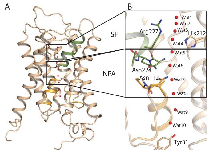
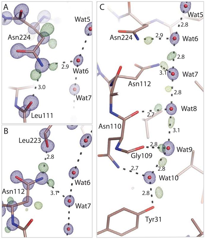
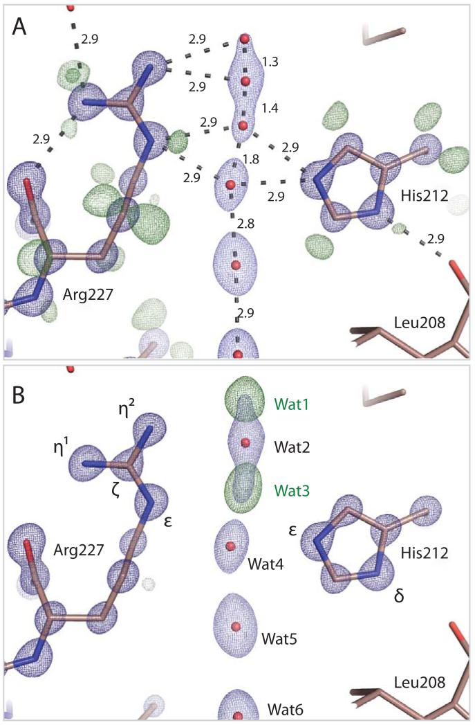
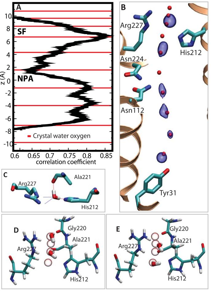
Aquaporins are membrane channels that facilitate the flow of water across biological membranes. Two conserved regions are central for selective function: the dual asparagine-proline-alanine (NPA) aquaporin signature motif and the aromatic and arginine selectivity filter (SF). Here, we present the crystal structure of a yeast aquaporin at 0.88 angstrom resolution. We visualize the H-bond donor interactions of the NPA motif's asparagine residues to passing water molecules; observe a polarized water-water H-bond configuration within the channel; assign the tautomeric states of the SF histidine and arginine residues; and observe four SF water positions too closely spaced to be simultaneously occupied. Strongly correlated movements break the connectivity of SF waters to other water molecules within the channel and prevent proton transport via a Grotthuss mechanism.


Figures

Figure 1
Figure 1
Fold of Aqy1. A, The six transmembrane helices and the pseudo seventh transmembrane helix formed by loops B (orange) and E (green). B, Water molecule positions within the channel (red spheres). The dual NPA aquaporin signature motif (lower box) and the SF (upper box) are highlighted.
Figure 2
Figure 2
Electron density within the NPA and cytoplasmic regions of the Aqy1. A, 2mFobs-DFcalc (blue, contoured at 4.3 e/Å3) and mFobs-DFcalc (green, contoured at 0.33 e/Å3) electron density associated with Asn224. B, 2mFobs-DFcalc (blue, contoured at 4.3 e/Å3) and mFobs-DFcalc (green, contoured at 0.33 e/Å3, yellow-green at 0.26 e/Å3) electron density associated with Asn112. Delocalized 2mFobs-DFcalc density connects the dual NPA asparagine Cγ and Oδ atoms whereas that associated with Nδ is more localized. Residual mFobs-DFcalc electron density indicates H-bond donor interactions to passing water molecules. C, 2mFobs-DFcalc electron density (blue, contoured at 4.3 e/Å3) illustrating the position of water molecules, and mFobs-DFcalc residual electron density (brown-green contoured at 0.59 e/Å3; dark green at 0.39 e/Å3; yellow-green 0.26-0.33 e/Å3; light green contoured at 0.15 e/Å3) indicating water H-bond interactions within the aquaporin channel.
Figure 3
Figure 3
Electron density within the Aqy1 SF. A, 2mFobs-DFcalc (dark blue, contoured at 4.3 e/Å3; light blue at 1.9 e/Å3) and residual mFobs-DFcalc (dark green, contoured at 0.42 e/Å3; light green contoured at 0.33 e/Å3) electron density associated with His212, Arg227 and water molecules within the SF. Atomic separations (Å) are indicated. Residual mFobs-DFcalc electron density reveals that Nδ of His212 is protonated whereas Nε is not. Connected 2mFobs-DFcalc electron density suggests that the Arg227 covalent bond from Cζ to Nη2 is preferentially conjugated. Four closely spaced water molecules are modeled within the SF with complementary occupancy (66% occupancy, positions 2 and 4; 34% occupancy, positions 1 and 3). B, mFobs-DFcalc omit electron density map calculated when Waters 1 and 3 are removed from the structural model (dark green, contoured at 0.65 e/Å3). Positive electron density features associated with these waters are the strongest within the channel.
Figure 4
Figure 4
Molecular dynamics simulations of water movements in Aqy1. A, Plot illustrating the correlation of movements of adjacent water molecules. Strongly correlated movements arise in the SF and in the cytoplasmic half of the channel. The position of oxygen atoms of crystallographic waters are indicated as red lines. B, Surface representation (blue) of the most probable positions of water molecules (averaged over the final 15 ns of a 20 ns trajectory) superimposed upon the crystallographic water positions (red spheres). C, Snapshot of a water molecule within the SF with all four H-bond interactions occupied. D and E, Snapshots corresponding to 1,3 and 2,4 water occupancy of the SF indicating the pair-wise movement of water molecules in this region. All four closely spaced SF crystallographic water positions are indicated as white spheres.

Comment in

  • Biochemistry. Watch water flow.
    Abramson J, Vartanian AS. Abramson J, et al. Science. 2013 Jun 14;340(6138):1294-5. doi: 10.1126/science.1239270. Science. 2013. PMID: 23766318 No abstract available.

References

    1. King LS, Kozono D, Agre P. From structure to disease: the evolving tale of aquaporin biology. Nat Rev Mol Cell Biol. 2004;5:687. - PubMed
    1. de Grotthuss CJT. Sur la décomposition de l’eau et des corps qu’elle tient en dissolution à l’aide de l’électricité galvanique. Ann. Chim. 1806;58:54.
    1. Marx D. Proton transfer 200 years after von Grotthuss: insights from ab initio simulations. Chemphyschem. 2006;7:1848. - PubMed
    1. Fu D, et al. Structure of a glycerol-conducting channel and the basis for its selectivity. Science. 2000;290:481. - PubMed
    1. Savage DF, Egea PF, Robles-Colmenares Y, O’Connell JD, 3rd, Stroud RM. Architecture and selectivity in aquaporins: 2.5 Å X-ray structure of aquaporin Z. PLoS Biol. 2003;1:E72. - PMC - PubMed

Publication types

Associated data