. 2012 Jan 17;109(3):811-6.
doi: 10.1073/pnas.1110270109. Epub 2011 Dec 29.

Structural basis for DNA recognition and loading into a viral packaging motor

Affiliations

Structural basis for DNA recognition and loading into a viral packaging motor

Carina R Büttner et al. Proc Natl Acad Sci U S A. .

Abstract

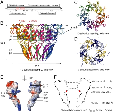
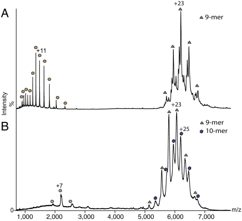
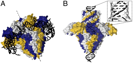
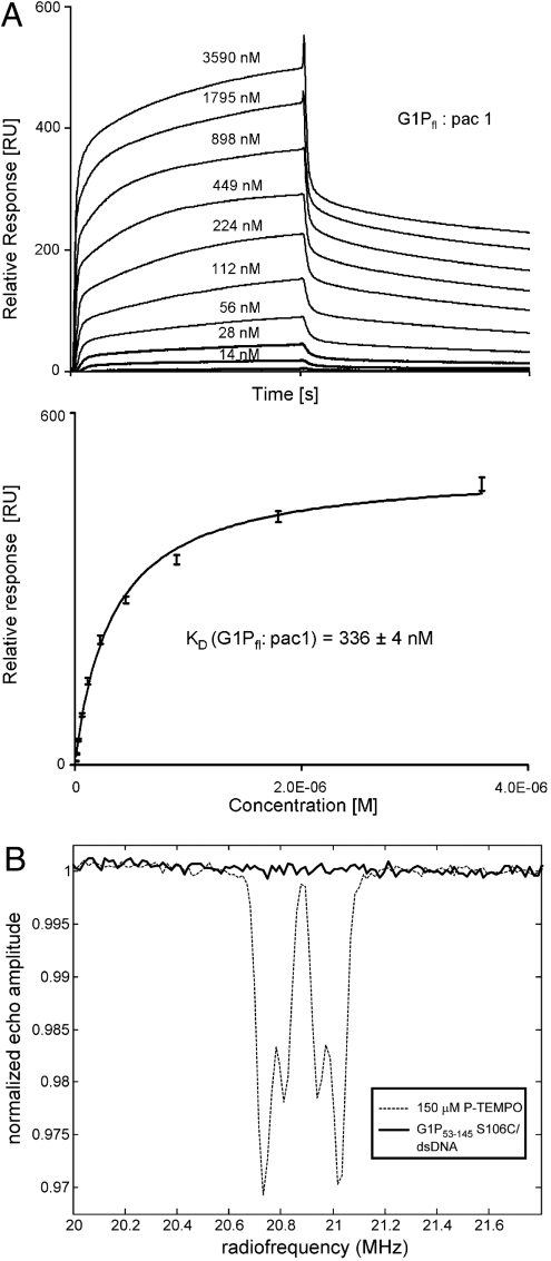
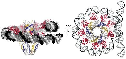
Genome packaging into preformed viral procapsids is driven by powerful molecular motors. The small terminase protein is essential for the initial recognition of viral DNA and regulates the motor's ATPase and nuclease activities during DNA translocation. The crystal structure of a full-length small terminase protein from the Siphoviridae bacteriophage SF6, comprising the N-terminal DNA binding, the oligomerization core, and the C-terminal β-barrel domains, reveals a nine-subunit circular assembly in which the DNA-binding domains are arranged around the oligomerization core in a highly flexible manner. Mass spectrometry analysis and four further crystal structures show that, although the full-length protein exclusively forms nine-subunit assemblies, protein constructs missing the C-terminal β-barrel form both nine-subunit and ten-subunit assemblies, indicating the importance of the C terminus for defining the oligomeric state. The mechanism by which a ring-shaped small terminase oligomer binds viral DNA has not previously been elucidated. Here, we probed binding in vitro by using EPR and surface plasmon resonance experiments, which indicated that interaction with DNA is mediated exclusively by the DNA-binding domains and suggested a nucleosome-like model in which DNA binds around the outside of the protein oligomer.


Conflict of interest statement

The authors declare no conflict of interest.

Figures

Fig. 1.
Fig. 1.
Structure of the oligomerization core domain (residues 53–120). (A) Domain organization showing location of the oligomerization core within the full-length protein. (B and C) Ribbon diagram of a ten-subunit assembly of G1P53–120 shown in two orthogonal views. The N and C termini as well as the secondary structure elements of a single subunit are labeled. “Linker” refers to residues 61–65 connecting α4 with the main body of the oligomerization core formed by helices α5 and α6. (D) Ribbon diagram of a nine-subunit assembly of the same construct, G1P53–120, shown in three alternating colors. (E) Electrostatic surface potential at subunit interface (ranging from -5, red, to +5 kT/e, blue) shown for two opposing subunits with the central axis vertical as in B. (F) Channel cross section indicating internal (van der Waals) diameters. The red stars indicate the position of the MTSSL in the S106C mutant used.
Fig. 2.
Fig. 2.
Structure of N-terminal deletion construct (residues 65–141) and full-length small terminase. (A) Ribbon diagram of the proteolytic fragment G1P65–141 containing the C-terminal β-barrel in addition to the main body of the oligomerization core domain shown with the central axis vertical, with individual subunits in alternating colors. The indicated van der Waals diameter of the β-barrel (9.8 Å) corresponds to the most constricted part of the channel. (B) Ribbon diagram of the full-length G1P shown along the axis with DBDs in red. Two DBDs of the asymmetric part (DBD-1 and DBD-2), that were observed in the electron density maps, are shown as ribbons, and the putative position of the third DBD (DBD-3) is depicted by dashed circles. (C) Models of full-length G1P shown with all DBDs either in DBD-1 (left) or DBD-2 (right) orientation. For clarity, DBDs of only five subunits are shown, with a 10-bp dsDNA-DBD complex modeled only for one subunit. The rotational and translational DBD movements derived from the normal mode analysis are indicated on the left. (D) Stereo figure comparing superposed individual subunits of small terminases from Siphoviridae SF6 phage (two adjacent subunits, red and pink), Podoviridae Sf6 phage (blue), and the putative small terminase from prophage phBC6A51 (yellow). The structures were aligned to fit the position of the channel axis, the first of the oligomerization α-helices and the C-terminal β-strand. Although the position of the two helices is conserved in the oligomer, the fold of the monomer differs in the Podoviridae Sf6 small terminase, where helix α6 occupies the same position as α6 of an adjacent subunit in the small terminases from the other two phages.
Fig. 3.
Fig. 3.
Mass spectrometry analysis. (A) Mass spectrum recorded for full-length G1P reveals a 9-mer centered at 6,000 m/z (green triangles). G1P has some minor C-terminal truncations (see Fig. S2), which arise from partial cleavage of residues 128–145. To resolve these peaks, increased acceleration voltage is applied leading to dissociation into monomers (low m/z of 1,000–2,000, brown circles). (B) Mass spectrum recorded for the construct G1P1–120 reveals the formation of both 9-mers (green triangles) and 10-mers (purple hexagon) in an approximately 1∶1 ratio. Gray circles correspond to monomers.
Fig. 4.
Fig. 4.
Potential models for the G1P-DNA complex. (A) DNA binding around the outside of the oligomer mediated by the DNA-binding domains. Each DBD in complex with a 10-bp oligonucleotide is reoriented so that DNA segments can form a continuous molecule. (B) DNA binding in the channel. The Inset demonstrates the match between the geometry of the C-terminal β-barrel and B-form DNA.
Fig. 5.
Fig. 5.
DNA binding. (A) SPR experiments for full-length G1P and the pac DNA recognition site comprising 428 bp (pac1). Equilibrium dissociation constant KD was estimated from a steady state analysis of three individual experiments. (B) EPR experiments. Mims ENDOR spectra for G1P53–145 S106C labeled with MTSSL at the inner channel surface as shown in Fig. 1F. The control experiment with 4-phosphonoxy-2,2,6,6-tetramethyl-piperidine-1-oxyl (dashed line) clearly shows phosphorous coupling, but no coupling was observed for the oligomer incubated with a 22-bp dsDNA (solid line).
Fig. 6.
Fig. 6.
Model of the small terminase-DNA complex during DNA recognition. Positions of DBDs were remodeled so that the vertical (channel) coordinate of the two extreme positions corresponds to coordinates observed in the crystal structure with all other DBDs occupying intermediate positions; orientation of each DBD was adjusted so that helix-turn-helix (HTH) motifs fit the helicoidal DNA, with approximately 34-Å spacing between adjacent HTH motifs.

References

    1. Rao VB, Feiss M. The bacteriophage DNA packaging motor. Annu Rev Genet. 2008;42:647–681. - PubMed
    1. Casjens SR. The DNA-packaging nanomotor of tailed bacteriophages. Nat Rev Microbiol. 2011;9:647–657. - PubMed
    1. Earnshaw WC, Casjens SR. DNA packaging by the double-stranded DNA bacteriophages. Cell. 1980;21:319–331. - PubMed
    1. Smith DE, et al. The bacteriophage ϕ29 portal motor can package DNA against a large internal force. Nature. 2001;413:748–752. - PubMed
    1. Camacho AG, Gual A, Lurz R, Tavares P, Alonso JC. Bacillus subtilis bacteriophage SPP1 DNA packaging motor requires terminase and portal proteins. J Biol Chem. 2003;278:23251–23259. - PubMed

Publication types

LinkOut - more resources