. 2015 May 1;290(18):11504-14.
doi: 10.1074/jbc.M115.637868. Epub 2015 Mar 17.

The class III cyclobutane pyrimidine dimer photolyase structure reveals a new antenna chromophore binding site and alternative photoreduction pathways

Affiliations

The class III cyclobutane pyrimidine dimer photolyase structure reveals a new antenna chromophore binding site and alternative photoreduction pathways

Patrick Scheerer et al. J Biol Chem. .

Abstract

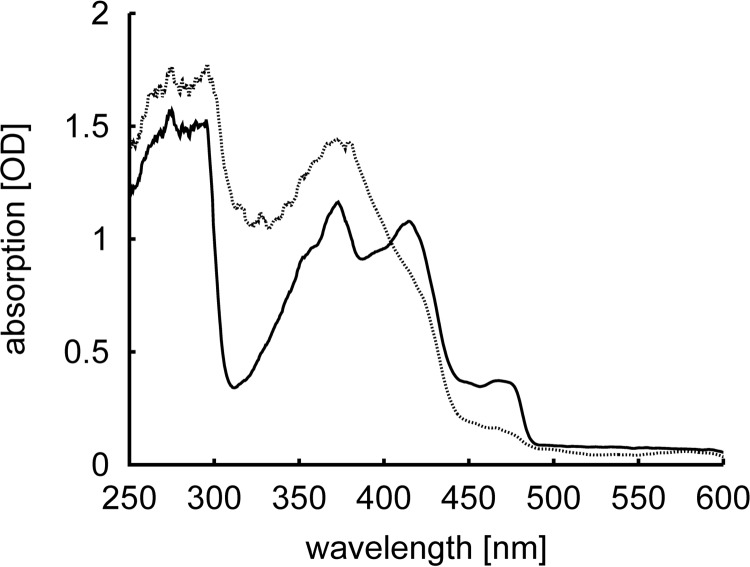
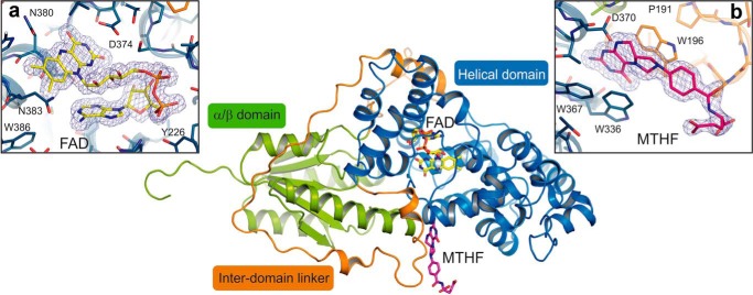
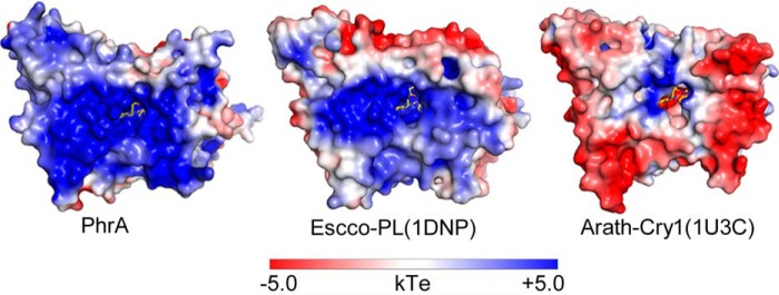
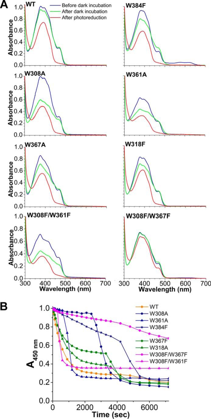
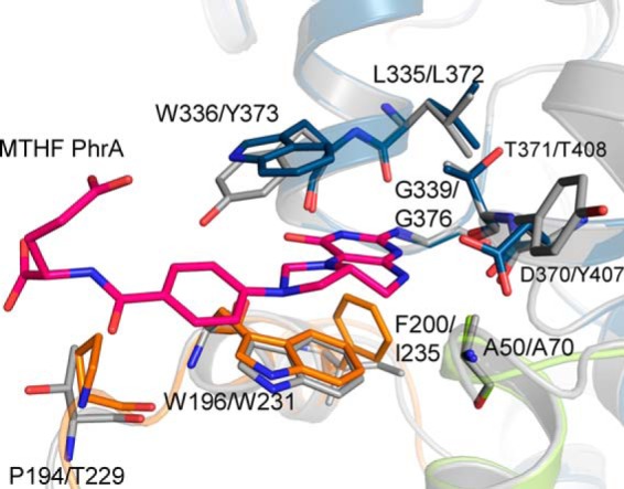
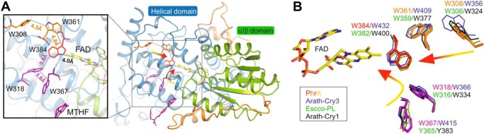
Photolyases are proteins with an FAD chromophore that repair UV-induced pyrimidine dimers on the DNA in a light-dependent manner. The cyclobutane pyrimidine dimer class III photolyases are structurally unknown but closely related to plant cryptochromes, which serve as blue-light photoreceptors. Here we present the crystal structure of a class III photolyase termed photolyase-related protein A (PhrA) of Agrobacterium tumefaciens at 1.67-Å resolution. PhrA contains 5,10-methenyltetrahydrofolate (MTHF) as an antenna chromophore with a unique binding site and mode. Two Trp residues play pivotal roles for stabilizing MTHF by a double π-stacking sandwich. Plant cryptochrome I forms a pocket at the same site that could accommodate MTHF or a similar molecule. The PhrA structure and mutant studies showed that electrons flow during FAD photoreduction proceeds via two Trp triads. The structural studies on PhrA give a clearer picture on the evolutionary transition from photolyase to photoreceptor.

Keywords: Cryptochrome; Crystal Structure; DNA Repair; Flavin Adenine Dinucleotide (FAD); Molecular Evolution; Mutagenesis in Vitro; Photobiology; Photoreceptor; Photoreduction; Site-directed Mutagenesis.


Figures

FIGURE 1.
FIGURE 1.
Phylogenetic tree with selected members of the CPF. Seven phylogenetic classes are indicated on the right and distinguished by different colors. CryP (written in black) appears separate and cannot be assigned to one of the classes. Abbreviations: Arath, A. thaliana; Agrtu, A. tumefaciens; Azoca, Azorhizobium caulinodans ORS 571; Braja, Bradyrhizobium japonicum USDA 110; Caucr, Caulobacter crescentus; Drome, D. melanogaster; Escco, E. coli; Homsa, Homo sapiens; Phatr, Phaeodactylum tricornutum; Oceal, Oceanocaulix alexandrii; Orysa, Oryza sativa; Rhosp, Rhodobacter sphaeroides 2.4.1; Rhopa, Rhodopseudomonas palustris; Sphsp, Sphingomonas sp. SKA58; Synel, Synechococcus elongatus PCC 6301, S6803: Synechocystis sp. PCC 6803; Theth, Thermus thermophilus HB8, Vibch, Vibrio cholerae; Xenla, Xenopus laevis; Cry, cryptochrome; DASH, DASH cryptochrome; PL64, (6-4)photolyase; Phr, photolyase; Plr, photolyase-related protein; CPD, cyclobutane pyrimidine dimer; FeS-BCP, iron-sulfur cluster containing bacterial cryptochromes and photolyases.
FIGURE 2.
FIGURE 2.
UV-visible absorption spectra measured in the PhrA crystal before (black straight line) and after (gray dotted line) x-ray exposure. Oxidized FAD has an absorption maximum at 450 nm, this absorbance is lost upon reduction.
FIGURE 3.
FIGURE 3.
Overall structure and cofactor arrangement of PhrA. The ribbon representation shows the α/β-domain (N-terminal domain; green) and the helical domain (catalytic domain; blue) connected by a long inter-domain linker (orange). The cofactors MTHF (colored in pink) and FAD (colored in yellow) are illustrated as sticks model. FAD/MTHF binding sites: inset a, FAD chromophore and inset b, MTHF ligand are shown as stick models with σA-weighted 2FoFc electron density maps contoured at 1.2 σ (blue mesh).
FIGURE 4.
FIGURE 4.
Electrostatic surface potentials of PhrA and comparison with E. coli photolyase and A. thaliana cryptochrome 1. Electrostatic surface potentials were calculated using the programs PyMol, APBS (67), and PDB code 2PQR with the non-linear Poisson-Boltzmann equation contoured at ± 5 kT/e. Negatively and positively charged surface areas are colored in red and blue, respectively.
FIGURE 5.
FIGURE 5.
Putative CPD lesion binding of PhrA. A, surface presentation of the Anani-PL crystal structure (PDB entry 1TEZ) and bound DNA with CPD; the six lesion-interacting amino acids are shown in blue, homologous amino acids of PhrA in green. B, enlarged view of the lesion contact, color code as in A; note the different orientation of Trp-386/Trp-392 in PhrA/Anani-PL, respectively. Inset, Trp-386 of PhrA and a potential rotamer of Trp-386 (brown) that could interact with the CPD lesion in the same manner as Trp-392 of Anani-PL.
FIGURE 6.
FIGURE 6.
MTHF binding sites of PhrA and other MTHF binding CPFs. A, MTHF chromophores of PhrA, E. coli CPD photolyase (Escco-PL, PDB entry 1DNP), and A. thaliana Cry3 (Arath-Cry3, PDB entry 2VTB). Distances between Asn-10 of MTHF and N5 of FAD are indicated. B, MTHF binding in PhrA. Potential hydrogen bonds or van der Waals contacts are shown as dashed lines. C, UV-visible spectra of wild type PhrA and the mutants W196A and W336A. D, putative MTHF cavity of plant cryptochrome, superposition of PhrA and A. thaliana Cry1 (Arath-Cry1, PDB entry 1U3C) with protein backbones drawn in green/blue/orange and gray, respectively. Relevant amino acids of Cry1 and the potential MTHF interacting amino acids of Arath-Cry1 are drawn as sticks; dashed lines indicated the same types of contacts as in C.
FIGURE 7.
FIGURE 7.
Photoreduction of PhrA. UV-visible spectral properties (A) and photoreduction of PhrA wild-type (WT) and mutated proteins (B). Absorption values at 450 nm were taken from UV-visible spectra measured at indicated time points upon onset of blue-light irradiation. For each protein, these values were normalized against the value measured at t = 0 min.
FIGURE 8.
FIGURE 8.
Proposed MTHF pocket of Phaeodactylum tricornutum CryP. The homology model, constructed with the Swissmodel server (16) using PhrA as template, is drawn in gray, the PhrA structure is drawn in green/blue/orange. Relevant amino acids are drawn as sticks and labeled; the first label refers to PhrA, the second to CryP.
FIGURE 9.
FIGURE 9.
Trp triads of PhrA and comparison with other CPF structures. A, two Trp triads in PhrA. The classical Trp triad is presented in red and orange, Trp of the alternative pathway are drawn in magenta. B, superposition of the residues of PhrA Trp triads (color code as in A) with Escco-PL (green lines), Arath-Cry1 (black lines), and Arath-Cry3 (blue lines).

References

    1. Sancar A. (2003) Structure and function of DNA photolyase and cryptochrome blue-light photoreceptors. Chem. Rev. 103, 2203–2237 - PubMed
    1. Müller M., Carell T. (2009) Structural biology of DNA photolyases and cryptochromes. Curr. Opin. Struct. Biol. 19, 277–285 - PubMed
    1. Chaves I., Pokorny R., Byrdin M., Hoang N., Ritz T., Brettel K., Essen L. O., van der Horst G. T., Batschauer A., Ahmad M. (2011) The cryptochromes: blue light photoreceptors in plants and animals. Annu. Rev. Plant Biol. 62, 335–364 - PubMed
    1. Oberpichler I., Pierik A. J., Wesslowski J., Pokorny R., Rosen R., Vugman M., Zhang F., Neubauer O., Ron E. Z., Batschauer A., Lamparter T. (2011) A photolyase-like protein from Agrobacterium tumefaciens with an iron-sulfur cluster. Plos One 6, e26775. - PMC - PubMed
    1. Selby C. P., Sancar A. (2006) A cryptochrome/photolyase class of enzymes with single-stranded DNA-specific photolyase activity. Proc. Natl. Acad. Sci. U.S.A. 103, 17696–17700 - PMC - PubMed

Publication types

MeSH terms

LinkOut - more resources