. 2015 Nov;24(11):1856-73.
doi: 10.1002/pro.2792. Epub 2015 Sep 8.

Weak conservation of structural features in the interfaces of homologous transient protein-protein complexes

Affiliations

Weak conservation of structural features in the interfaces of homologous transient protein-protein complexes

Govindarajan Sudha et al. Protein Sci. 2015 Nov.

Abstract

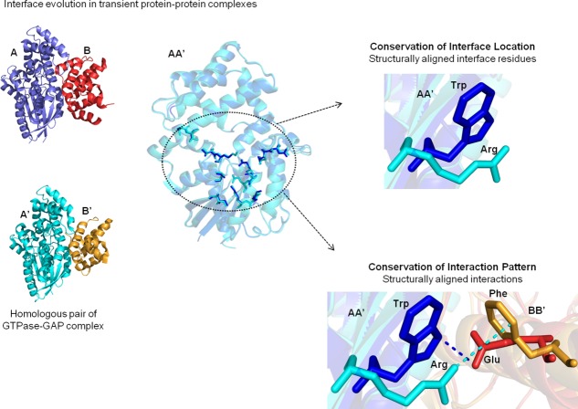
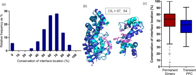
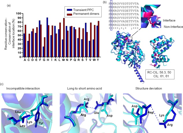
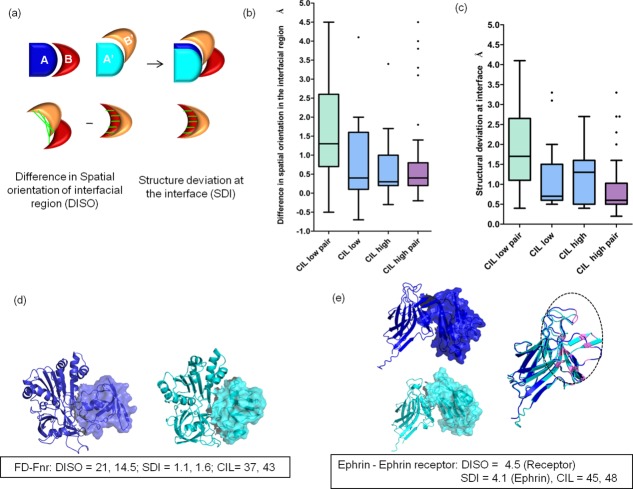
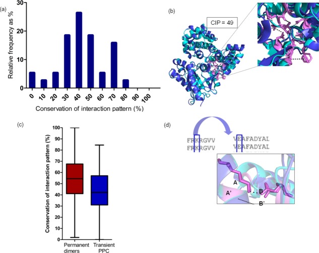
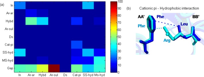
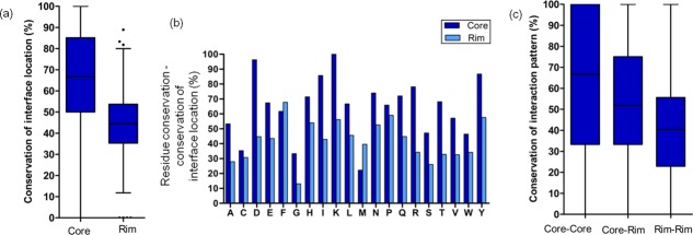
Residue types at the interface of protein-protein complexes (PPCs) are known to be reasonably well conserved. However, we show, using a dataset of known 3-D structures of homologous transient PPCs, that the 3-D location of interfacial residues and their interaction patterns are only moderately and poorly conserved, respectively. Another surprising observation is that a residue at the interface that is conserved is not necessarily in the interface in the homolog. Such differences in homologous complexes are manifested by substitution of the residues that are spatially proximal to the conserved residue and structural differences at the interfaces as well as differences in spatial orientations of the interacting proteins. Conservation of interface location and the interaction pattern at the core of the interfaces is higher than at the periphery of the interface patch. Extents of variability of various structural features reported here for homologous transient PPCs are higher than the variation in homologous permanent homomers. Our findings suggest that straightforward extrapolation of interfacial nature and inter-residue interaction patterns from template to target could lead to serious errors in the modeled complex structure. Understanding the evolution of interfaces provides insights to improve comparative modeling of PPC structures.

Keywords: evolution of protein complexes; interfaces of protein complexes; protein-protein interactions; structure of protein complexes; transient protein complexes.


Figures

Figure 1
Figure 1
Extent of conservation of interaction interfaces in homologous transient protein–protein complexes. Homologous pairs of transient protein–protein complexes (PPCs) (A:B and A′:B′) are shown, where A is homologous to A′ and B is homologous to B′. Interface residues in AA′ that are structurally equivalent are circled. Structurally aligned interface residues are said to have conserved interface location (CIL), whereas structurally aligned interactions are said to have conserved interaction pattern (CIP). The interfacial features do not consider the residue type or the nature of interactions.
Figure 2
Figure 2
Extent of conservation of interface location (CIL) in homologous transient PPCs. (a) Histogram shows distribution of CIL scores for homologous pairs of transient PPCs. (b) Homologous pair of guanine nucleotide-binding protein G (o) subunit alpha (regulator of G-protein signaling 16) and guanine nucleotide-binding protein G (k) subunit alpha (regulator of G-protein signaling 10) (2ihb, 3c7k) is colored in dark and light blue. Both the homologous subunits show moderate CIL score. Residues with CIL are shown as pink sticks. All the protein structure figures in this work are generated using PyMOL. (c) Box plot distribution for CIL in homologous permanent dimers and transient PPC. The lines from bottom specify the minimum, 25th percentile, median, 75th percentile, and maximum for all the box plots in this work. The outliers of the distribution are represented as dots.
Figure 3
Figure 3
Extent of conserved residue types in homologous subunits of protein complexes with conserved interface location. (a) Extent of structurally equivalent conserved residue type to be at interface (residue conservation-conservation of interface location, RC-CIL) for each of the 20 residue types for homologous transient PPC and permanent homodimers are shown. (b) Conserved residue type in a multiple sequence alignment of homologs need not imply that all the conserved residues should lie at the interface. An example of structurally equivalent arginine residues from homologous subunits shows one arginine at the interface (pink) and the other arginine not in the interface (magenta). The extent of structurally equivalent, conserved residue types to be at the interface (RC-CIL = 58.35%, 50%) and conservation of interface location (CIL = 61%, 61%) have been calculated for the homologous subunits of GTPase-GAP complex. (c) In spite of residue types being conserved, interface location is lost in homologous protein complex (dark and light blue) by different ways.
Figure 4
Figure 4
Reasons for non-conservation of interface location in homologous transient PPC. (a) Differences in spatial orientation of the interfacial region (DISO) is calculated by subtracting the RMSD of the interfacial region of homologous subunits (red and orange subunits) in their interfacial region with respect to the structurally aligned proteins (dark and light blue subunits) with RMSD for the interfacial region for the isolated homologous subunits. Structural differences at the interface (SDI) are calculated by calculating the RMSD for the interfacial region in isolated homologous subunits. Distance between the structurally equivalent interface residues are shown as green lines. (b) Box plot distribution for DISO scores for CIL low pair (both the homologous subunits of protein complex have low CIL score (30%–60%)), CIL low–CIL high pair (one homologous subunit has a low CIL score, whereas the other homologous subunit has high CIL score (>60%)), and CIL high pair (both the homologous subunits of protein complex have high CIL score). (c) Box plot distribution for SDI scores for homologous subunits of transient PPC is shown. (d) Homologous pair of ferredoxin: ferredoxin-NADP+ reductase (PDB id: 1ewy, 1gaq) shows drastic DISO. Changes in the spatial orientation are shown by homologous subunits, which are represented as surface. High DISO scores and low CIL scores for both the homologous subunits of the protein complex are observed. (e) Homologous pair of Ephrin-Ephrin receptor complex showing high DISO score for the ephrin receptors and high SDI for the ephrin molecules results in low CIL score (45%, 48%). Changes in the spatial orientation in homologous ephrin receptors are represented as surface. Structure deviation at the interface in the homologous ephrin molecules are highlighted in the circled region.
Figure 5
Figure 5
Extent of conservation of interaction pattern in homologous transient PPC. (a) Histogram of conservation of interaction pattern (CIP) scores for homologous pairs of transient PPC is colored in blue. (b) Homologous pair of guanine nucleotide-binding protein G (o) subunit alpha (regulator of G-protein signaling 16) and guanine nucleotide-binding protein G (k) subunit alpha (regulator of G-protein signaling 10) (2ihb, 3c7k) is colored in dark and light blue. Residues with CIP are shown as pink sticks, and common interactions are shown as dotted lines. (c) Box plot distribution for CIP in homologous permanent dimers and transient PPC. (d) Residue–residue interaction in a particular transient PPC (K-E) has their residue type conserved in the other homolog. This mostly results in residue–residue interactions being maintained in the other homolog.
Figure 6
Figure 6
Interaction type substitutions for residues with conserved interaction pattern in homologous transient protein–protein complexes. (a) Interaction type substitution for residues with CIP in homologous pairs of transient PPC is shown. Various interaction types include hydrophobic (hybd), disulfide (Ds), side chain–side chain hydrogen bonding (SS-hyd), main chain–side chain hydrogen bonding (MS-hyd), ionic (In), aromatic–aromatic (Ar-Ar), aromatic–sulfur (Ar-sul), and cation–pi (Cat-pi). Gap denotes non-conservation of interaction pattern. (b) Cation–pi (Phe-Arg) interaction is getting substituted to hydrophobic interaction (Phe-Leu) in residues with CIP in homologous pairs of transient PPC. Residues with CIP are shown as sticks, and the common interactions are shown as dotted lines.
Figure 7
Figure 7
Extent of conservation of interface location and interaction pattern in core and rim interface residues. (a) Extent of core and rim interface residues to show conserved interface location. (b) Extent of core and rim residues that have conserved residue type and conserved interface location have been shown for all the 20 residue types. (c) Extent of core–core, core–rim, and rim–rim interactions to show conserved interaction pattern.

References

    1. Janin J, Bahadur RP, Chakrabarti P. Protein-protein interaction and quaternary structure. Q Rev Biophys. 2008;41:133–180. - PubMed
    1. Keskin O, Gursoy A, Ma B, Nussinov R. Principles of protein–protein interactions: what are the preferred ways for proteins to interact? Chem Rev. 2008;108:1225–1244. - PubMed
    1. Bahadur RP, Chakrabarti P, Rodier F, Janin J. Dissecting subunit interfaces in homodimeric proteins. Proteins. 2003;53:708–719. - PubMed
    1. Nooren IM, Thornton JM. Diversity of protein-protein interactions. EMBO J. 2003;22:3486–3492. - PMC - PubMed
    1. Ozbabacan SE, Engin HB, Gursoy A, Keskin O. Transient protein-protein interactions. Protein Eng Des Sel. 2011;24:635–648. - PubMed

Publication types

Substances

Associated data

LinkOut - more resources