. 2018 Nov 16;293(46):17705-17715.
doi: 10.1074/jbc.RA118.003830. Epub 2018 Sep 21.

Chromophorylation of cyanobacteriochrome Slr1393 from Synechocystis sp. PCC 6803 is regulated by protein Slr2111 through allosteric interaction

Affiliations

Chromophorylation of cyanobacteriochrome Slr1393 from Synechocystis sp. PCC 6803 is regulated by protein Slr2111 through allosteric interaction

Qi He et al. J Biol Chem. .

Abstract

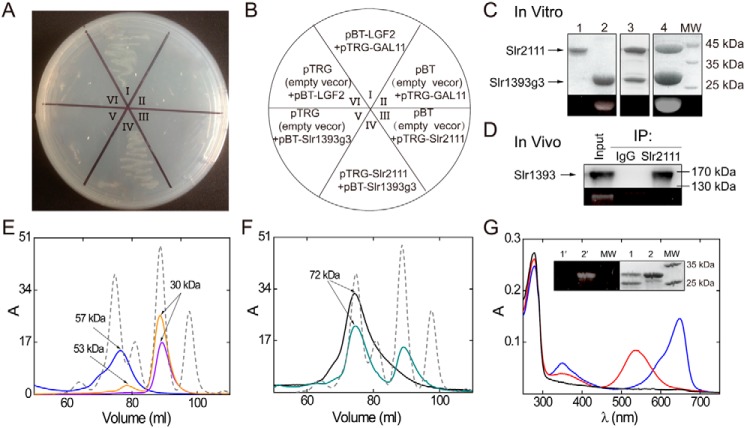
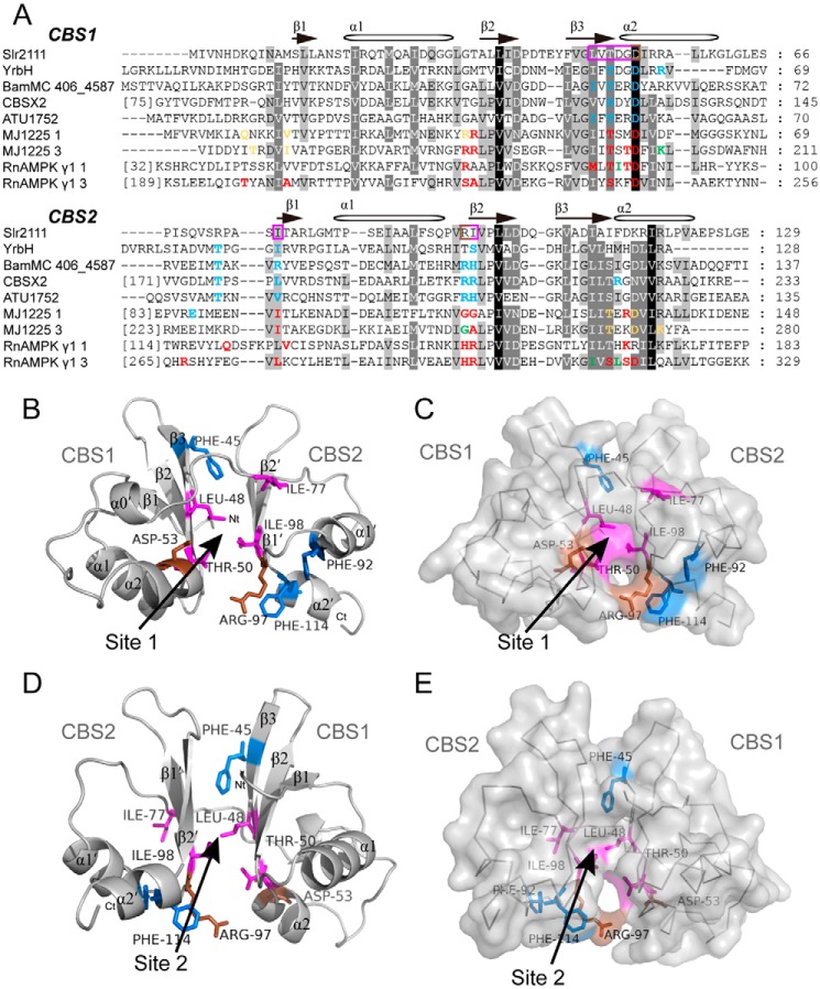
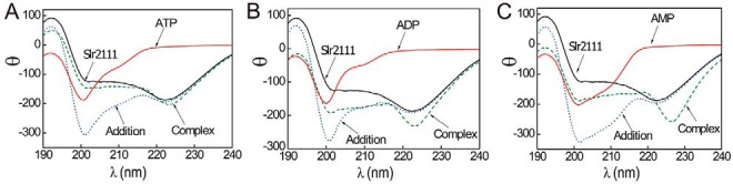
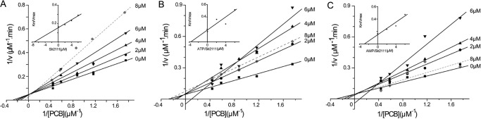
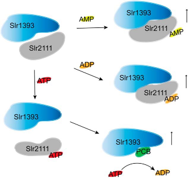
Cyanobacteriochromes (CBCRs) are photochromic proteins in cyanobacteria that act as photosensors. CBCRs bind bilins as chromophores and sense nearly the entire visible spectrum of light, but the regulation of the chromophorylation of CBCRs is unknown. Slr1393 from Synechocystis sp. PCC 6803 is a CBCR containing three consecutive GAF (cGMP phosphodiesterase, adenylyl cyclase, and FhlA protein) domains, of which only the third one (Slr1393g3) can be phycocyanobilin-chromophorylated. The protein Slr2111 from Synechocystis sp. PCC 6803 includes a cystathionine β-synthase (CBS) domain pair of an as yet unknown function at its N terminus. CBS domains are often characterized as sensors of cellular energy status by binding nucleotides. In this work, we demonstrate that Slr2111 strongly interacts with Slr1393 in vivo and in vitro, which generates a complex in a 1:1 molar ratio. This tight interaction inhibits the chromophorylation of Slr1393g3, even if the chromophore is present. Instead, the complex stability and thereby the chromophorylation of Slr1393 are regulated by the binding of nucleotides (ATP, ADP, AMP) to the CBS domains of Slr2111 with varying affinities. It is demonstrated that residues Asp-53 and Arg-97 of Slr2111 are involved in nucleotide binding. While ATP binds to Slr2111, the association between the two proteins gets weaker and chromophorylation of Slr1393 are enabled. In contrast, AMP binding to Slr2111 leads to a stronger association, thereby inhibiting the chromophorylation. It is concluded that Slr2111 acts as a sensor of the cellular energy status that regulates the chromophorylation of Slr1393 and thereby its function as a light-driven histidine kinase.

Keywords: adenosine-binding protein; co-immunoprecipitation; cyanobacteria; cystathionine beta-synthase domain; photoreceptor; photosynthesis; photosynthetic pigment; phototransduction; phycocyanobilin; protein-protein interaction; regulation.


Conflict of interest statement

The authors declare that they have no conflicts of interest with the contents of this article

Figures

Figure 1.
Figure 1.
Protein interaction of Slr2111 and Slr1393g3. A, protein interaction demonstrated by the BacterioMatch II Two-hybrid System. The reporter strain containing pBT-Slr1393g3 + pTRG-Slr2111 (A, IV) grew well in the presence of 3-AT and streptomycin. B, the wheel shows the details of the plate of A. The reporter strains II, VI, III, and V acted as negative controls, and I acted as the positive control. C, SDS-PAGE control of pulldown assay results; lane 1, elution fractions of GST-Slr2111 (43 kDa); lane 2, PCB-His6tag-Slr1393g3 (24 kDa); lane 3, elution fraction of PCB-His6tag-Slr1393g3 and GST-Slr2111 on GST affinity chromatography; lane 4, elution fraction of PCB-His6tag-Slr1393g3 and GST-Slr2111 on His6-tag affinity column. The bottom panel shows the Zn2+-induced fluorescence of the chromoprotein band on SDS-PAGE. D, co-immunoprecipitations of Slr1393 and Slr2111. Input: lysate from Synechocystis. Slr2111 antibodies were bound to protein A beads and used for immunoprecipitation from total Synechocystis sp. PCC 6803 soluble proteins. Immunoprecipitates with anti-Slr2111 antibodies or control immunoglobulin G (IgG) was assayed with SDS-PAGE without reducing agent and then Western blotted with anti-Slr1393. The bottom panel shows the Zn2+-induced fluorescence of the chromoprotein band on SDS-PAGE. E, gel filtration chromatography profiles of His6tag-Slr2111 (blue line), PCB-His6tag-Slr1393g3 (orange line), His6tag-Slr1393g3 (violet line), using a Superdex 200 column; elution buffer: KPB (20 mm, pH 7.4) containing NaCl (200 mm) (see “Materials and methods”). Blue line, peak corresponds to Slr2111 as dimer (57 kDa, calculated 45 kDa). Violet line, peak corresponds to Slr1393g3 as monomer (30 kDa, calculated 24 kDa). Orange line, two peaks correspond to PCB-Slr1393g3 as monomer (30 kDa, calculated 24 kDa) and dimer (53 kDa, calculated 48 kDa), respectively. Molecular markers (peaks from left to right of gray dashed line) were 66, 45, 29, and 12.4 kDa; F, the complex of Slr2111 and Slr1393g3 in a 1:1 ratio elutes with an apparent mass of 72 kDa (black line), and the mixture of Slr2111 and PCB-Slr1393g3 in a 1:1 ratio elutes with the respective apparent mass of 72 and 30 kDa (green line) under the same experimental conditions as in E. Gel chromatography identifies the complex of Slr2111 and Slr1393g3 as a mixture of heterodimer (72 kDa, calculated 47 kDa) and heterotetramer (72 kDa, calculated 94 kDa). G, absorption spectrum (black line) of the early peak of the green line in F purified by gel chromatography, absorption after irradiation with 530 nm (Z state, blue line), and 650-nm light (E state, red line) of the later peak of the green line in F were measured. Inset G, SDS-PAGE of the early peak (lane 1) and later peak (lane 2) of the green line in F; left panel 1′ and 2′ show the Zn2+-induced fluorescence of the chromoprotein band on SDS-PAGE.
Figure 2.
Figure 2.
Sequence alignment of CBS domains and model building of Slr2111. A, sequence alignments were performed by GeneDoc. Nucleotide-binding sites are colored: green, amino acids binding ATP; blue, amino acids binding AMP; yellow, amino acids binding ADP; amino acids binding more than two kinds of nucleotides are shown in red. Possible binding sites of Slr2111 are highlighted in a violet frame. B–E, simulation of the Slr2111 structure (10–123 aa). The three-dimensional model was obtained from the SWISS-MODEL server, and a putative signal-transduction protein with CBS domains from B. ambifaria MC40–6 (PDB code 4fry) was used as reference structure (see “Materials and methods” for details). Side chains of aromatic residues are indicated for three phenylalanine residues (Phe-45, Phe-92, and Phe-114) (blue). Leu-48, Thr-50, Ile-77, and Ile-98 are shown in violet, Asp-53 and Arg-97 are shown in brown. D and E depicts the structure after rotation around 180 degrees of B and C, respectively. Two possible binding sites of adenine nucleotides are indicated. Image was generated in PyMOL.
Figure 3.
Figure 3.
Far-UV CD of Slr2111 complexed with adenosine ligands. A, spectra of isolated Slr2111 (black line), the isolated ATP (red line), the addition of the spectra of both isolated species (blue dotted line), and the complex formed by equimolar (10 μm) amounts of both biomolecules (green dashed line). B, spectra of isolated Slr2111, the isolated ADP, the complex Slr2111/ADP, and the addition of both spectra of the isolated biomolecules (color coding as in A). C, spectra of isolated Slr2111, the isolated AMP, the complex Slr2111/AMP, and the addition of both spectra of the isolated biomolecules (color coding as in A). Experiments were carried out at 25 °C.
Figure 4.
Figure 4.
Lineweaver-Burk plots in the inhibition of Slr1393g3 by (A) Slr2111, (B) Slr2111/ATP, and (C) Slr2111/AMP. Numbers on the different lines indicate Slr2111 concentrations (0–8 μm). PCB was used at concentrations of 0, 0.5, 0.8, 1.1, 1.7, and 2.3 μm. The slopes of the solid lines by inhibitors (0–6 μm) was used to calculate for Ki with a secondary plot (insets A–C). Competitive inhibition was observed with respect to Slr2111, Slr2111/ATP, and Slr2111/AMP, the calculated Ki values were 7.3, 3.8, and 2.9 μm, respectively.
Figure 5.
Figure 5.
Model for interaction of Slr1393g3 with Slr2111 that regulates the chromophorylation of Slr1393. As levels of AMP and ADP increase in cells, Slr2111 binds free AMP and ADP, which leads to a conformation change of Slr2111 and is more convenient for binding Slr1393g3, so inhibiting PCB-chromophorylation of Slr1393. As the ATP level increased, the complex of Slr2111 and Slr1393 is more liable to dissociation, free Slr1393 is subject to PCB-chromophorylation, so cascading the photoactivation of HK and signal transmission. It should be noted that the HK converts ATP to ADP, thereby decreasing the level of ATP, which leads to the decrease of the chromophorylation of Slr1393g3. The vertical up arrows indicate that the yields of the products increase.

References

    1. Ikeuchi M., and Ishizuka T. (2008) Cyanobacteriochromes: a new superfamily of tetrapyrrole-binding photoreceptors in cyanobacteria. Photochem. Photobiol. Sci. 7, 1159–1167 10.1039/b802660m - DOI - PubMed
    1. Rockwell N. C., Martin S. S., Feoktistova K., and Lagarias J. C. (2011) Diverse two-cysteine photocycles in phytochromes and cyanobacteriochromes. Proc. Natl. Acad. Sci. U.S.A. 108, 11854–11859 10.1073/pnas.1107844108 - DOI - PMC - PubMed
    1. Fushimi K., Nakajima T., Aono Y., Yamamoto T., Ni-Ni W., Ikeuchi M., Sato M., and Narikawa R. (2016) Photoconversion and fluorescence properties of a red/green-type cyanobacteriochrome AM1_C0023g2 that binds not only phycocyanobilin but also biliverdin. Front. Microbiol. 7, 588 - PMC - PubMed
    1. Narikawa R., Fukushima Y., Ishizuka T., Itoh S., and Ikeuchi M. (2008) A novel photoactive GAF domain of cyanobacteriochrome AnPixJ that shows reversible green/red photoconversion. J. Mol. Biol. 380, 844–855 10.1016/j.jmb.2008.05.035 - DOI - PubMed
    1. Chen Y., Zhang J., Luo J., Tu J. M., Zeng X. L., Xie J., Zhou M., Zhao J. Q., Scheer H., and Zhao K. H. (2012) Photophysical diversity of two novel cyanobacteriochromes with phycocyanobilin chromophores: photochemistry and dark reversion kinetics. FEBS J. 279, 40–54 10.1111/j.1742-4658.2011.08397.x - DOI - PubMed

Publication types

MeSH terms

LinkOut - more resources