. 2012 Jun 13;31(12):2824-32.
doi: 10.1038/emboj.2012.107. Epub 2012 Apr 20.

Csy4 relies on an unusual catalytic dyad to position and cleave CRISPR RNA

Affiliations

Csy4 relies on an unusual catalytic dyad to position and cleave CRISPR RNA

Rachel E Haurwitz et al. EMBO J. .

Abstract

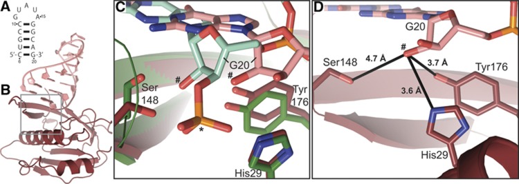
CRISPR-Cas adaptive immune systems protect prokaryotes against foreign genetic elements. crRNAs derived from CRISPR loci base pair with complementary nucleic acids, leading to their destruction. In Pseudomonas aeruginosa, crRNA biogenesis requires the endoribonuclease Csy4, which binds and cleaves the repetitive sequence of the CRISPR transcript. Biochemical assays and three co-crystal structures of wild-type and mutant Csy4/RNA complexes reveal a substrate positioning and cleavage mechanism in which a histidine deprotonates the ribosyl 2'-hydroxyl pinned in place by a serine, leading to nucleophilic attack on the scissile phosphate. The active site catalytic dyad lacks a general acid to protonate the leaving group and positively charged residues to stabilize the transition state, explaining why the observed catalytic rate constant is ∼10(4)-fold slower than that of RNase A. We show that this RNA cleavage step is essential for assembly of the Csy protein-crRNA complex that facilitates target recognition. Considering that Csy4 recognizes a single cellular substrate and sequesters the cleavage product, evolutionary pressure has likely selected for substrate specificity and high-affinity crRNA interactions at the expense of rapid cleavage kinetics.


Conflict of interest statement

The authors declare that they have no conflict of interest.

Figures

Figure 1
Figure 1
Amino acid contributions to catalysis. (A) Csy4 active site from Csy4/substrate complex (PDB ID 2XLK). Active site residues are shown in stick format and the scissile phosphate is marked with an asterisk. The hydrogen bonds of the base pair between nucleotides C6 and dG20 are shown as dashed lines. (B) Representative single-turnover cleavage assays with wild-type and mutant Csy4. No protein (NP) controls shown at left. (C) Single-turnover cleavage analysis of wild-type and mutant Csy4. Data plotted are average of triplicate experiments and error bars represent the standard error of the mean (s.e.m.). Solid lines represent fits to an exponential equation. (D) pH-rate profile for wild-type and H29K Csy4. Rapid cleavage kinetics above pH 9.5 for wild-type Csy4 prevented accurate determination of the rate. Each data point is an average of three independent experiments and error bars represent the s.e.m. Data were fit according to the equation described in the Materials and Methods.
Figure 2
Figure 2
Crystal structure of Csy4/product RNA complex at 2.0 Å resolution. (A) Shown at left is the substrate RNA used to generate the protein/RNA complex. Cleavage by Csy4 (purple arrow) produces the product RNA (right) present in the crystal structure. Gray lettering denotes nucleotides for which there was no corresponding electron density and therefore could not be modeled. (B) Overall structure of Csy4S22C (dark green) bound to product RNA (light green). Electron density was well-defined for all 187 amino acids of Csy4 and 16 of the 19 nucleotides in the product RNA. (C) Detailed view of the Csy4 active site (gray box, in B). The 2′-hydroxyl nucleophile is marked with a pound sign and the scissile phosphate is marked with an asterisk. RNA/protein hydrogen-bonding interactions are marked with dashes.
Figure 3
Figure 3
Crystal structure of Csy4S148A/RNA complex at 2.6 Å resolution. (A) Shown at left is the substrate RNA used to generate the protein/RNA complex. Cleavage by Csy4 (purple arrow) produces product RNA (right). Because of the slow cleavage rate of the S148A mutant, crystals likely contained a mixed population of substrate and product RNAs. (B) Overall structure of Csy4S148A (dark purple) and RNA (light purple). 153/187 amino acids and 14/15 nucleotides could be modeled into the electron density. The amino acids composing the arginine-rich helix are among those for which there is little to no electron density. (C) Superposition and close-up of product complex (green) and S148A complex (purple) active sites (gray box, in B). The double-headed black arrow highlights the 3.2 Å change in 2′-hydroxyl location between the two structures. The two 2′-hydroxyl nucleophiles are labeled with pound signs and the scissile phosphates are indicated with an asterisk.
Figure 4
Figure 4
Crystal structure of Csy4/RNA minimal complex at 2.3 Å resolution. (A) The stem-loop RNA used for co-crystallography lacks a 3′-phosphate. (B) Overall structure of Csy4 (dark red) and stem-loop RNA (pink). 151/187 amino acids and all 15 RNA nucleotides could be modeled into the electron density. Electron density for the active site loop is severely broken, and a dashed line indicates its approximate location. There is no electron density for the arginine-rich helix. (C) Superposition and detailed view of product complex (green) and minimal complex (red) active sites (gray box, in B). The scissile phosphate belonging to the product complex is marked with an asterisk and the two 2′-hydroxyl nucleophiles are marked with pound signs. (D) Magnified view of the minimal complex active site. Black lines indicate the distances between active site residues and the 2′-hydroxyl nucleophile.
Figure 5
Figure 5
Csy4 cleavage of pre-crRNA is required for Csy complex formation. (A) Schematic depicting pre-crRNA cleavage by Csy4 and formation of the Csy CRISPR ribonucleoprotein (crRNP) complex. The CRISPR repeat and spacer sequence are in black and green, respectively. Cleavage sites are denoted with purple arrows. (B) Superose 6 gel filtration column elution profiles of affinity-purified Csy1, Csy2, His6-Csy3, and pre-crRNA co-expressed with wild-type (blue) or H29A (red) Csy4. (C) Coomassie blue-stained 12% SDS–PAGE showing protein components of the superose 6 fractions for wild-type (lane 1) and H29A (lanes 2–4, as noted in B) Csy4 co-expression assays. (D) SYBR Gold-stained 15% denaturing PAGE showing phenol:chloroform extracted nucleic acids from superose 6 fractions (from B).

References

    1. Adams PD, Afonine PV, Bunkoczi G, Chen VB, Davis IW, Echols N, Headd JJ, Hung LW, Kapral GJ, Grosse-Kunstleve RW, McCoy AJ, Moriarty NW, Oeffner R, Read RJ, Richardson DC, Richardson JS, Terwilliger TC, Zwart PH (2010) PHENIX: a comprehensive Python-based system for macromolecular structure solution. Acta Crystallogr D Biol Crystallogr 66: 213–221 - PMC - PubMed
    1. Al-Attar S, Westra ER, van der Oost J, Brouns SJ (2011) Clustered regularly interspaced short palindromic repeats (CRISPRs): the hallmark of an ingenious antiviral defense mechanism in prokaryotes. Biol Chem 392: 277–289 - PubMed
    1. Barrangou R, Fremaux C, Deveau H, Richards M, Boyaval P, Moineau S, Romero DA, Horvath P (2007) CRISPR provides acquired resistance against viruses in prokaryotes. Science 315: 1709–1712 - PubMed
    1. Bolotin A, Ouinquis B, Sorokin A, Ehrlich SD (2005) Clustered regularly interspaced short palindrome repeats (CRISPRs) have spacers of extrachromosomal origin. Microbiology-Sgm 151: 2551–2561 - PubMed
    1. Brouns SJ, Jore MM, Lundgren M, Westra ER, Slijkhuis RJ, Snijders AP, Dickman MJ, Makarova KS, Koonin EV, van der Oost J (2008) Small CRISPR RNAs guide antiviral defense in prokaryotes. Science 321: 960–964 - PMC - PubMed

Publication types

MeSH terms