. 2012 Dec;24(12):5074-88.
doi: 10.1105/tpc.112.104422. Epub 2012 Dec 18.

Crystal structure of rice importin-α and structural basis of its interaction with plant-specific nuclear localization signals

Affiliations

Crystal structure of rice importin-α and structural basis of its interaction with plant-specific nuclear localization signals

Chiung-Wen Chang et al. Plant Cell. 2012 Dec.

Abstract

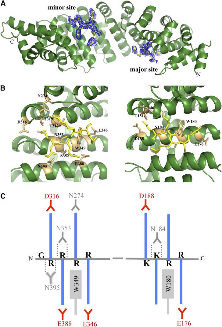
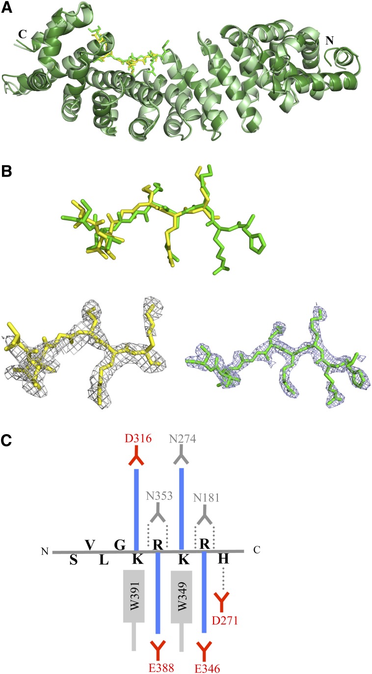
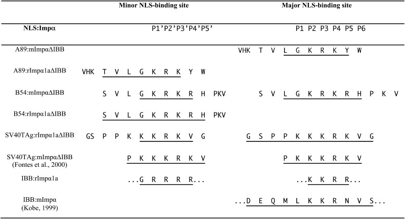
In the classical nucleocytoplasmic import pathway, nuclear localization signals (NLSs) in cargo proteins are recognized by the import receptor importin-α. Importin-α has two separate NLS binding sites (the major and the minor site), both of which recognize positively charged amino acid clusters in NLSs. Little is known about the molecular basis of the unique features of the classical nuclear import pathway in plants. We determined the crystal structure of rice (Oryza sativa) importin-α1a at 2-Å resolution. The structure reveals that the autoinhibitory mechanism mediated by the importin-β binding domain of importin-α operates in plants, with NLS-mimicking sequences binding to both minor and major NLS binding sites. Consistent with yeast and mammalian proteins, rice importin-α binds the prototypical NLS from simian virus 40 large T-antigen preferentially at the major NLS binding site. We show that two NLSs, previously described as plant specific, bind to and are functional with plant, mammalian, and yeast importin-α proteins but interact with rice importin-α more strongly. The crystal structures of their complexes with rice importin-α show that they bind to the minor NLS binding site. By contrast, the crystal structures of their complexes with mouse (Mus musculus) importin-α show preferential binding to the major NLS binding site. Our results reveal the molecular basis of a number of features of the classical nuclear transport pathway specific to plants.


Figures

Figure 1.
Figure 1.
The Structure of Full-Length rImpα1a. (A) The structure of rImpα1a comprises 10 ARM repeats (green, cartoon representation) and two NLS-like sequences from the N-terminal IBB domain (shown in yellow stick representation, superimposed with simulated annealing omit electron density map contoured at 2σ). (B) The NLS-like sequences from the IBB domain (G25RRRR29 and K47KRR50 in yellow stick representation) interact with the labeled residues (in orange stick representation) from the minor (left) and major NLS binding sites (right), respectively, of rImpα1a. (C) Schematic illustration of the main interactions between the IBB domain and rImp1a. The basic side chains (blue color) of Lys or Arg from NLS-like segments form salt bridges and electrostatic interactions with acid residues (red). The aliphatic portions of the basic chains interact with the Trp and Asn (in gray). The hydrogen bonds (dotted lines) are formed between Asn residues (in gray color) and the main-chain amides.
Figure 2.
Figure 2.
Structure and Sequence Conservation in Impα Proteins. (A) Structure-based alignment of mImpα, yImpα, and rImpα1a sequences (including the alignment of ARM repeats). Red highlights conserved residues; blue indicates insertions and deletions; green indicates the NLS-like segments. (B) Superposition of full-length rice (green), mouse (blue; Kobe 1999), and yeast (magenta; from export complex structure; Matsuura and Stewart 2004) Impα proteins (ribbon representation). (C) Sequence conservation of plant Impα proteins mapped onto the surface of rImpα1a (Consurf; Ashkenazy et al., 2010), based on the alignment of Impα proteins from Nicotiana benthamiana, Arabidopsis lyrata ssp lyrata, O. sativa ssp japonica, Trifolium pratense, Capsicum annuum, A. thaliana, and Z. mays. Red indicates conserved, while blue indicates variable.
Figure 3.
Figure 3.
A89 and B54 NLS Peptides Can Be Translocated into the Nucleus Using Impα Proteins from Different Organisms. Nuclear import assays were performed in permeabilized human (HEp-2) cells. The cargo proteins (GST-GFP) fused to various NLS peptides (A89, B54, and SV40TAg) were transported into the nucleus by mouse Impβ and mImpα, yImpα, and rImpα1a. (A) The left panel shows localization of GFP; the middle panel shows localization of Texas-red dextran (70 kD; magenta), showing the condition of the nuclear membranes; and the right panel is the merged image with 4′,6-diamidino-2-phenylindole staining indicating the location of the nuclei (blue). (B) Import assay control experiments. The first three panels show that a cargo without NLS (GST-GFP) is not translocated into the nucleus by Impα:mImpβ. In the last three panels, GST-GFP-SV40TAgNLS import by mImpα1a:mImpβ failed due to the lack of an ATP generating system (no ATP added), lack of transporter proteins (no mImpα:Impβ), or addition of wheat germ agglutinin (WGA). (C) The images show that the majority of the cells have intact nuclear membranes as indicated by the accumulation of Texas-red dextran (magenta) in the cytoplasm and GST-GFP-SV40TAgNLS (green) being translocated to the nucleus. We counted 2166 nuclei, and <4% of those were permeable to Texas-red dextran.
Figure 4.
Figure 4.
A89 and B54 NLS Peptides Bind to the Minor NLS Binding Site of rImpα1aΔIBB. (A) Superposition of rImpα1aΔIBB:A89 (light green/yellow) and rImpα1aΔIBB:B54 (dark green/green) complex structures (cartoon representation, NLS peptides in stick representation). (B) Simulated annealing omit electron density maps (contoured at 2σ; gray mesh) superimposed onto the structure of A89 (top, yellow) and B54 (bottom, green) NLSs (stick representation). (C) Schematic illustration of the interactions between B54NLS and rImp1a. The basic side chains (blue color) of Lys or Arg form salt bridges and electrostatic interactions with acid residues (red). The aliphatic portions of the basic chains interact with the Trp and Asn (in gray). The hydrogen bonds (dotted lines) are formed between Asn residues (in gray color) and the main-chain amides.
Figure 5.
Figure 5.
NLS Binding to Specific Binding Pockets in rImpα1a and mImpα Proteins, Based on Structural Information. The NLS-like sequences are aligned based on the interaction with Impα binding sites. The residues visible in the electron density map are underlined.
Figure 6.
Figure 6.
Differential Binding of Plant-Specific NLSs to rImpα1a and mImpα. (A) B54NLS:rImpα1aΔIBB complex. Top: The C-terminal residues of B54NLS (SVL3 in green stick representation) bound to rImpα1aΔIBB (gray cartoon representation, contact residues in gray stick representation). Bottom: Top view of the pocket (in gray surface representation) that accommodates Leu3 from the peptide. (B) Superposition of the mImpαΔIBB:B54NLS and rImpα1aΔIBB:B54NLS structures. Top: mImpαΔIBB:B54NLS (light blue, cartoon representation, B54NLS omitted) and rImpα1aΔIBB:B54NLS (green, stick representation, rImpα1a omitted) in a view analogous to (A). Leu-3 from the peptide shows steric hindrance with Thr-402 (in magenta stick representation) from mImpαΔIBB. Bottom: Top view of the pocket (in light blue surface representation). Thr-402 is shown in red surface representation.

References

    1. Adams P.D., et al. (2010). PHENIX: A comprehensive Python-based system for macromolecular structure solution. Acta Crystallogr. D Biol. Crystallogr. 66: 213–221 - PMC - PubMed
    1. Ashburner M., et al. ; The Gene Ontology Consortium (2000). Gene ontology: Tool for the unification of biology. Nat. Genet. 25: 25–29 - PMC - PubMed
    1. Ashkenazy H., Erez E., Martz E., Pupko T., Ben-Tal N. (2010). ConSurf 2010: Calculating evolutionary conservation in sequence and structure of proteins and nucleic acids. Nucleic Acids Res. 38(Web Server issue): W529–W533. - PMC - PubMed
    1. Bethke G., Unthan T., Uhrig J.F., Pöschl Y., Gust A.A., Scheel D., Lee J. (2009). Flg22 regulates the release of an ethylene response factor substrate from MAP kinase 6 in Arabidopsis thaliana via ethylene signaling. Proc. Natl. Acad. Sci. USA 106: 8067–8072 - PMC - PubMed
    1. Catimel B., Teh T., Fontes M.R., Jennings I.G., Jans D.A., Howlett G.J., Nice E.C., Kobe B. (2001). Biophysical characterization of interactions involving importin-alpha during nuclear import. J. Biol. Chem. 276: 34189–34198 - PubMed

MeSH terms

LinkOut - more resources