. 2013 Jun 18;110(25):10183-8.
doi: 10.1073/pnas.1300547110. Epub 2013 Jun 3.

High-resolution structures of the IgM Fc domains reveal principles of its hexamer formation

Affiliations

High-resolution structures of the IgM Fc domains reveal principles of its hexamer formation

Roger Müller et al. Proc Natl Acad Sci U S A. .

Abstract

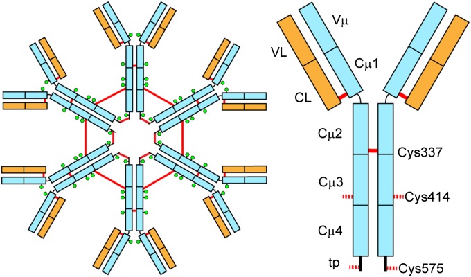
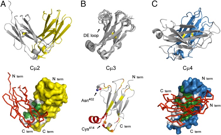
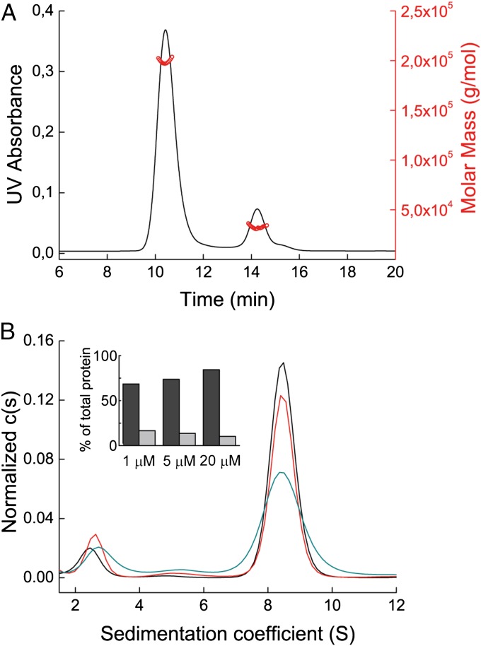
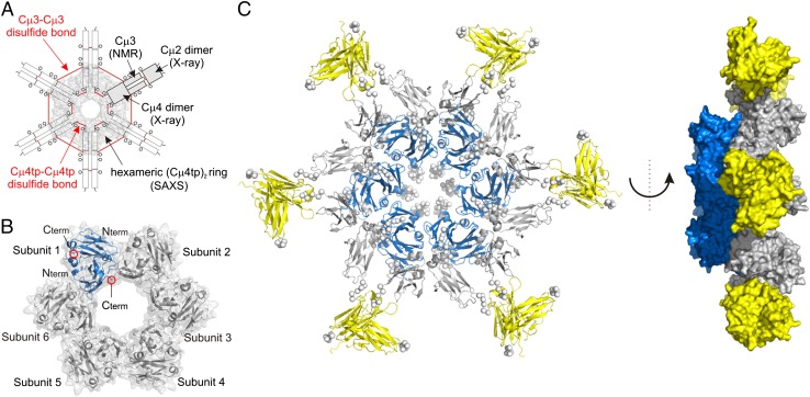
IgM is the first antibody produced during the humoral immune response. Despite its fundamental role in the immune system, IgM is structurally only poorly described. In this work we used X-ray crystallography and NMR spectroscopy to determine the atomic structures of the constant IgM Fc domains (Cµ2, Cµ3, and Cµ4) and to address their roles in IgM oligomerization. Although the isolated domains share the typical Ig fold, they differ substantially in dimerization properties and quaternary contacts. Unexpectedly, the Cµ4 domain and its C-terminal tail piece are responsible and sufficient for the specific polymerization of Cµ4 dimers into covalently linked hexamers of dimers. Based on small angle X-ray scattering data, we present a model of the ring-shaped Cµ4 structure, which reveals the principles of IgM oligomerization.

Keywords: antibody oligomerization; dimer interfaces; hybrid approach.


Conflict of interest statement

The authors declare no conflict of interest.

Figures

Fig. 1.
Fig. 1.
Schematic view of hexameric IgM (Left) and a single subunit (Right). Heavy chains are depicted in light blue and light chains in orange. Red lines represent intersubunit disulfide bridges and the green hexagons show glycosylation sites. The cysteine residues that form interdomain disulfide bridges between the Cµ2 domains (C337) and covalently link the IgM hexamer in Cµ3 (Cys414) and the C-terminal tp (C575) are depicted.
Fig. 2.
Fig. 2.
Structures of the individual IgM Fc domains. (A) Cartoon representation of the crystal structure of the Cµ2 domain (Upper, Protein Data Bank ID code 4JVU), the arrangement of the two molecules in the crystallographic asymmetric unit forming a stable dimer. Inter- and intramolecular disulfide bonds are shown in stick representation. The surface of one Cµ2 domain (chain A, yellow), showing the contact area with the second Cµ2 domain (chain B, red), is represented as Cα-trace (Lower). (B) Solution structure of the Cµ3 domain (Protein Data Bank ID code 4BA8). Superposition of the 10 lowest-energy NMR structures are shown as cartoon (Upper). The highly flexible DE loop is labeled. Chemical shift perturbations in the Cµ3 domain, when bound to the Cµ4 domain, are shown (Lower). The color scale is as follows: yellow, 0.025–0.05 ppm; orange, 0.05–0.1 ppm; and red, >0.1 ppm. The positions for the interdomain disulfide bridge (Cys414) and the glycosylation site (Asn402) are labeled and shown in stick representation. (C) Cartoon representation of the crystal structure the Cµ4 domain (Upper, Protein Data Bank ID code 4JVW). Intramolecular disulfide bonds are shown in stick representation. The surface of one Cµ4 domain (chain A, blue), showing the contact area with the second Cµ4 domain (chain B, red), is represented as Cα-trace (Lower). Color coding for the interfaces: gray, hydrophilic contact residues; green, hydrophobic contact residues.
Fig. 3.
Fig. 3.
Characterization of the Cμ4tp oligomer. (A) SEC MALS profile of the Cµ4tp oligomer. UV absorbance is shown in black, and calculated masses in red. (B) Cµ4tp oligomer characterized by aUC SV runs at 20 μM (black), 5 μM (red), and 1 μM (cyan). The histogram shows the percentages of hexamer of dimers (dark gray) and dimers (light gray) of the total protein in solution.
Fig. 4.
Fig. 4.
SAXS analysis of Cµ4tp and hexameric IgM Fc modeling. (A) Scheme of the IgM Fc hexamer with the information used for model building is indicated. (B) Structural model of the Cµ4tp hexamer of dimers (Protein Data Bank ID code 4BLE). The hexameric subunits and N/C termini of the Cµ4 dimer structure are annotated. One single Cµ4tp dimer is shown in blue. (C) Cartoon representation of the IgM Fc hexamer model (Left). A side view of the Fc hexamer is given (Right), showing that the inner core (Cµ4) is protruding out of the plane defined by the Cµ2 and Cµ3 domains. Cµ2 dimers are shown in yellow, Cµ3 in gray, and the Cµ4 core ring in blue. The termini and linker residues are represented as Cα dummy atoms by the program CORAL and are shown as spheres.

References

    1. Wannemuehler MJ, Galvin JE. Bacterial immunogens and protective immunity in swine. Vet Immunol Immunopathol. 1994;43(1-3):117–126. - PubMed
    1. Ohanian SH, Schlager SI. Humoral immune killing of nucleated cells: mechanisms of complement-mediated attack and target cell defense. Crit Rev Immunol. 1981;1(3):165–209. - PubMed
    1. Boes M. Role of natural and immune IgM antibodies in immune responses. Mol Immunol. 2000;37(18):1141–1149. - PubMed
    1. Thomas HI, Morgan-Capner P. The avidity of specific IgM detected in primary rubella and reinfection. Epidemiol Infect. 1990;104(3):489–497. - PMC - PubMed
    1. Taylor B, et al. C1q binding properties of monomer and polymer forms of mouse IgM mu-chain variants. Pro544Gly and Pro434Ala. J Immunol. 1994;153(11):5303–5313. - PubMed

Publication types