. 2012 Nov 20;109(47):19256-61.
doi: 10.1073/pnas.1210938109. Epub 2012 Nov 6.

Watching a signaling protein function in real time via 100-ps time-resolved Laue crystallography

Affiliations

Watching a signaling protein function in real time via 100-ps time-resolved Laue crystallography

Friedrich Schotte et al. Proc Natl Acad Sci U S A. .

Abstract

To understand how signaling proteins function, it is crucial to know the time-ordered sequence of events that lead to the signaling state. We recently developed on the BioCARS 14-IDB beamline at the Advanced Photon Source the infrastructure required to characterize structural changes in protein crystals with near-atomic spatial resolution and 150-ps time resolution, and have used this capability to track the reversible photocycle of photoactive yellow protein (PYP) following trans-to-cis photoisomerization of its p-coumaric acid (pCA) chromophore over 10 decades of time. The first of four major intermediates characterized in this study is highly contorted, with the pCA carbonyl rotated nearly 90° out of the plane of the phenolate. A hydrogen bond between the pCA carbonyl and the Cys69 backbone constrains the chromophore in this unusual twisted conformation. Density functional theory calculations confirm that this structure is chemically plausible and corresponds to a strained cis intermediate. This unique structure is short-lived (∼600 ps), has not been observed in prior cryocrystallography experiments, and is the progenitor of intermediates characterized in previous nanosecond time-resolved Laue crystallography studies. The structural transitions unveiled during the PYP photocycle include trans/cis isomerization, the breaking and making of hydrogen bonds, formation/relaxation of strain, and gated water penetration into the interior of the protein. This mechanistically detailed, near-atomic resolution description of the complete PYP photocycle provides a framework for understanding signal transduction in proteins, and for assessing and validating theoretical/computational approaches in protein biophysics.


Conflict of interest statement

The authors declare no conflict of interest.

Figures

Fig. 1.
Fig. 1.
PYP structure and photocycle. (A) Surface rendering (gray) of PYP (PDB ID code 2ZOH) in the pG state with backbone structure (ribbon) and atomic rendering of pCA and its hydrogen-bonding partners. Arrows point to the C2=C3 double bond (pCA) and the Cα atoms of key residues. Hydrogen bonds are indicated with dashed blue lines. Arg52 is stabilized in its “closed” state via hydrogen bonds to the protein backbone. [rendered with VMD (www.ks.uiuc.edu/Research/vmd/)] (B) The C2=C3 double bond in pCA is trans in its ground state; it absorbs blue light and gives PYP its yellow color. Photoisomerization from trans to cis triggers the PYP photocycle. (C) The PYP photocycle is labeled and color-coded according to intermediates characterized in this study. Intermediates colored gray and connected by dashed lines were not observed in this study, but have been characterized or implicated in prior studies.
Fig. 2.
Fig. 2.
Pump-probe geometry used to acquire time-resolved diffraction snapshots. The PYP crystal is sealed in a thin-walled glass capillary. Because the laser penetration depth in PYP is shallow, an orthogonal pump-probe geometry is used in which the top edge of the protein crystal is positioned at the top edge of the focused X-ray pulse. This geometry ensures optimal overlap between the laser and X-ray illuminated volumes of the crystal. The protein crystal acts as a monochromator with various line spacings (d) and diffracts different X-ray colors (λ) in different directions (θ) according to Bragg’s law (λ = 2d sinθ). Approximately 3,000 spots are found in each time-resolved diffraction image. The spots in this figure are annotated according to integrated photons (spot dimension) and X-ray wavelength (spot color).
Fig. 3.
Fig. 3.
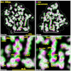
Front and side views of time-resolved structural changes recorded 100 ps after photoexcitation of PYP. (Lower) Expanded, annotated view of Upper Insets. The ground-state electron density map is colored magenta, and the 100-ps map is colored green. Where magenta and green overlap, the electron density blends to white. The magenta-to-green color gradient unveils the direction of atomic motion; large-amplitude displacements are indicated with yellow arrows. The stick models correspond to refined structures for the ground state (pG) and the first intermediate (pR0). Hydrogen bonds to the pCA chromophore are indicated as dotted lines.
Fig. 4.
Fig. 4.
Time-resolved population of transient intermediates and their structures. (A) Kinetic model used to account for structural changes spanning 10 decades; the arrows are labeled with the inverse of the globally refined rate constants. Half the population short-circuits to the ground state during the pR2-to-pB0 transition. (B) Time-dependent populations of each intermediate in the PYP photocycle: theoretical population predicted by the kinetic model (solid lines) and least-squares contributions from the four electron-density base maps (filled circles). (C–F) Structures for pR0, pR1, pR2, and pB0 intermediates. (Left and Center Left) Electron-density base maps were derived from global analysis and phased according to their refined structures (front and side views; see Fig. S3 for comparison with maps generated using ground-state phases). (Center Right) Refined structures of pCA intermediates and their hydrogen-bonding partners. To highlight the structural changes leading to the corresponding intermediate, they are overlaid with a semitransparent structure (gray) of the preceding state. (Right) Color-coded rendering of the protein backbone according to Cα displacement relative to pG, as indicated by the scale (rendered with VMD).

References

    1. Eaton WA, Henry ER, Hofrichter J. Nanosecond crystallographic snapshots of protein structural changes. Science. 1996;274(5293):1631–1632. - PubMed
    1. Srajer V, et al. Photolysis of the carbon monoxide complex of myoglobin: Nanosecond time-resolved crystallography. Science. 1996;274(5293):1726–1729. - PubMed
    1. Schotte F, et al. Watching a protein as it functions with 150-ps time-resolved x-ray crystallography. Science. 2003;300(5627):1944–1947. - PubMed
    1. Meyer TE. Isolation and characterization of soluble cytochromes, ferredoxins and other chromophoric proteins from the halophilic phototrophic bacterium Ectothiorhodospira halophila. Biochim Biophys Acta. 1985;806(1):175–183. - PubMed
    1. Hustede E, Liebergesell M, Schlegel HG. The photophobic response of various sulfur and nonsulfur purple bacteria. Photochem Photobiol. 1989;50(6):809–815.

Publication types

Substances