. 2013 Dec 13;288(50):35714-25.
doi: 10.1074/jbc.M113.510461. Epub 2013 Oct 30.

Structure of the cyanobacterial phytochrome 2 photosensor implies a tryptophan switch for phytochrome signaling

Affiliations

Structure of the cyanobacterial phytochrome 2 photosensor implies a tryptophan switch for phytochrome signaling

Katrin Anders et al. J Biol Chem. .

Abstract

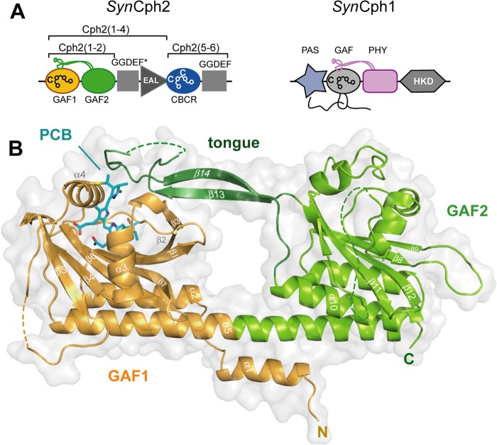
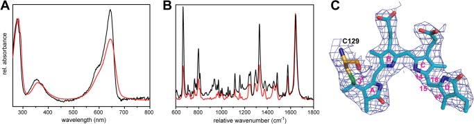
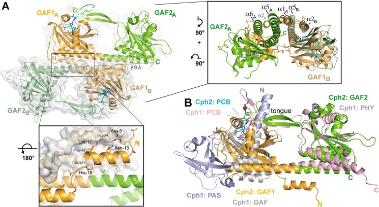
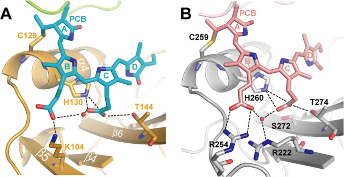
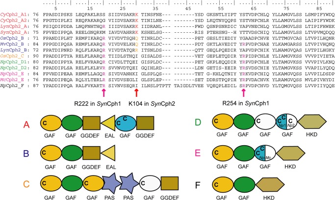
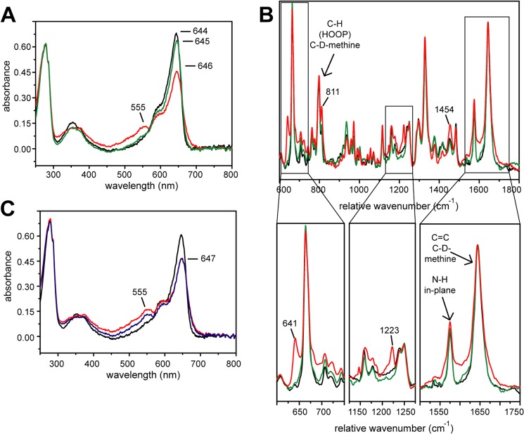
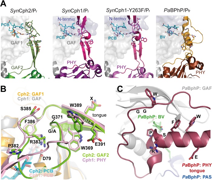
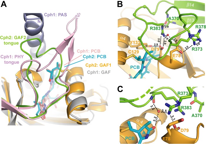
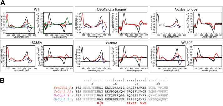
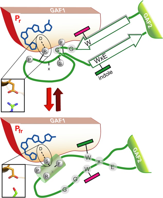
Phytochromes are highly versatile photoreceptors, which occur ubiquitously in plants as well as in many light-responsive microorganisms. Here, photosynthetic cyanobacteria utilize up to three different phytochrome architectures, where only the plant-like and the single-domain cyanobacteriochromes are structurally characterized so far. Cph2 represents a third group in Synechocystis species and affects their capability of phototaxis by controlling c-di-GMP synthesis and degradation. The 2.6-Å crystal structure of its red/far-red responsive photosensory module in the Pr state reveals a tandem-GAF bidomain that lacks the figure-of-eight knot of the plant/cph1 subfamily. Its covalently attached phycocyanobilin chromophore adopts a highly tilted ZZZssa conformation with a novel set of interactions between its propionates and the GAF1 domain. The tongue-like protrusion from the GAF2 domain interacts with the GAF1-bound chromophore via its conserved PRXSF, WXE, and W(G/A)G motifs. Mutagenesis showed that the integrity of the tongue is indispensable for Pr → Pfr photoconversion and involves a swap of the motifs' tryptophans within the tongue-GAF1 interface. This "Trp switch" is supposed to be a crucial element for the photochromicity of all multidomain phytochromes.

Keywords: Biliprotein; Cyanobacteria; Photochromicity; Phytochrome; Protein Conformation; Red Light Photoreceptor; Signal Transduction; Signaling; Structural Biology; c-di-GMP Signaling.


Figures

FIGURE 1.
FIGURE 1.
Structure of the SynCph2(1–2) photosensor. A, domain organization of SynCph1 and SynCph2. The tongue-like regions protruding from the GAF2 and PHY domains are shown in green and purple, respectively; the knot in SynCph1 as black lines. SynCph1 and SynCph2 differ in their effector domains: a histidine kinase domain (HKD) and c-di-GMP turnover (GGDEF and EAL) and regulatory (GGDEF*) domains. B, crystal structure of the SynCph2(1–2) module. GAF2 (green) contains a tongue-like region (dark green) for sealing the PCB (cyan)-binding pocket. The GAF1 domain is in orange.
FIGURE 2.
FIGURE 2.
Spectral characteristics of SynCph2(1–2) in soluble and crystallized forms. A, UV-visible absorbance spectra of SynCph2(1–2) in solution at room temperature (red curve) and a crystal (black) at 100 K in the Pr conformation. B, Raman spectra of the protein in frozen solution (red curve) at 100 K and in crystalline form (black) at 100 K. C, omit electron density of the PCB chromophore and its covalent attachment to Cys129 (contouring level 1.0 σ).
FIGURE 3.
FIGURE 3.
Quaternary structure of the knotless SynCph2(1–2) photosensor and comparison to SynCph1. A, the antiparallel dimer of SynCph2(1–2). The distance between C-terminal residues His421 (molecule A) and Ile418 (molecule B) is 89 Å; lower inlet, the N-terminal helix forms part of the dimer interface; inlet on the right side, perpendicular view on the SynCph2(1–2) dimer. The interface between the monomers is built of a helix bundle composed of the linker α-helix and shorter helices, especially the N-terminal helix. B, structural superposition of SynCph2 and SynCph1 (r.m.s. deviation 2.64 Å for 249 Cα atoms); the PAS, GAF, and PHY domains of SynCph1 are depicted in blue, gray, and pale red, respectively.
FIGURE 4.
FIGURE 4.
Close-up view of the chromophore binding sites of the SynCph2(1–2) (A) and SynCph1 (B) photosensor. The PCB chromophore within SynCph2(1–2) is structurally distorted compared with the almost planar A-, B-, and C-rings of SynCph1. The hydrogen bond network of the B- and C-ring propionates is indicated with dashed lines.
FIGURE 5.
FIGURE 5.
Multiple sequence alignment of Cph2-like Group II phytochromes. GAF1 sequences forming the knot/propionate binding site region. Abbreviations: SynCph2_A (organism: Synechocystis sp. PCC 6803; GenBank entry NP_442466.1; sequence identity: 100%), CyCph2_A1 (Cyanothece sp. PCC 7424; YP_002378924.1; 38%), CyCph2_A2 (Cyanothece sp. PCC 7822; YP_003886347.1; 39%), LynCph2_A (Lyngbya sp. PCC 8106; WP_009783799.1; 33%), OsCph2_B (Oscillatoria sp. PCC 6506; WP_007353933.1; 36%), MvCph2_B (Microcoleus vaginatus FGP-2; WP_006623727.1; 34%), LynCph2_B (Lyngbya sp. PCC 8106; WP_009783371.1; 33%), OsCph2_C (Oscillatoria sp. PCC 6506; WP_007357482.1; 30%), NpCph2_D1 (N. punctiforme PCC 73102; YP_001870049.1; 31%), NpCph2_D2 (N. punctiforme PCC 73102; YP_001868068.1; 33%), NpCph2_E (N. punctiforme PCC 73102; YP_001868577.1; 34%), McCph2_E (Microcoleus chthonoplastes PCC 7420; WP_006100748.1; 31%), NpCph2_F (N. punctiforme PCC 73102; YP_001865364.1; 31%). Pairwise sequence homologies to SynCph2(1–2) were performed with the BLOSUM62 exchange matrix. Letters at the end of the abbreviation describe the domain architecture of the proteins described below: C indicates conserved cysteines; green, GAF domain of the “PHY”-type; blue, GAF domain of the “CBCR”-type; white, GAF domain with sequence similarities with “CBCR”-type but with one or no conserved cysteine. Mixed colors, gene-dependent domain organization as indicated by the organism abbreviation.
FIGURE 6.
FIGURE 6.
Temporary radiation effect on SynCph2(1–2). Black, no x-ray; red, after x-ray; green, after annealing; blue, 60 s after exposure. A, UV-visible absorbance spectra of a crystal before x-ray, after 60 s exposure (ID 14-1 (ESRF, Grenoble), 100% transmission; flux: ∼4.2 × 10¹⁰ photons/s (approximately ∼1.4 × 10¹⁰ photons/s reached the crystal)), and after 3 s of annealing in the dark (annealing process was not complete). B, Raman spectra of a crystal before x-ray, after 30 min x-ray irradiation (ID 14–1 (ESRF, Grenoble), 100% transmission, 4-s exposure time per image, 350 images, 0.5° oscillation range; flux: ∼4.8 × 10¹⁰ photons/s) and after 4 s of annealing in the dark; below: partial scale-up of the Raman spectra. C, UV-visible absorbance spectra of a crystal before x-ray, after exposition for 60 s, and 60 s after exposure.
FIGURE 7.
FIGURE 7.
The tongue-like region of SynCph2(1–2). A, structure and interactions between the tongues and the chromophore-bearing GAF domains of phytochromes. B, comparison of the tongue regions of SynCph2 and SynCph1, superimposition of the GAF domains (r.m.s. deviation = 1.095 Å for 125 Cα); GAF1 and GAF2 of SynCph2 are displayed in orange and green; PAS, GAF, and PHY of SynCph1 in blue, gray, and pale red, respectively. The chromophores are shown in cyan (Cph2) and red (Cph1). The positions of the PRXSF, W(G/A)G, and WXE motifs in the tongue regions are conserved between SynCph1 and SynCph2, the residues are numbered along the SynCph2 count. C, tongue region of PaBphP (PDB code 3NHQ), the PAS (blue), GAF (gray) and PHY (red) domains and biliverdin in green. In contrast to SynCph2 and SynCph1 the tongue region consists of an extended loop region and α-helical elements. The spatial positions of the conserved motifs differ in PaBphP. The orientation of the tongue region correlates to that of SynCph2.
FIGURE 8.
FIGURE 8.
The tip of the tongue region of SynCph2. A, comparison of the photosensory modules of SynCph2 and SynCph1 at the tongue-GAF1 interface, color-coding corresponds to Fig. 7. B, the tongue region seals the PCB-binding pocket. In molecule B the whole tongue is defined by electron density. Only Arg383 of the PRXSF motif (spheres) is depicted as stick representation. Hydrogen bond distances are given in Å. C, chain A shows that the tip of the tongue is rather motile as several residues are missing and Arg373 adopts a different conformation.
FIGURE 9.
FIGURE 9.
Mutants and hybrids of the SynCph2 tongue region. A, UV-visible absorbance (left) and CD spectra (right) of SynCph2(1–2) wild type and mutants after far-red (Pr-state, black line) and red light illumination (red). Difference spectra (APrAPhotoequilibrium) are shown in blue. Green curves in the wild type spectra are calculated for pure Pfr (10). The partial lack of a wild type-like Pfr CD spectrum of Oscillatoria tongue is due to its instability. B, multiple sequence alignment of Group II phytochromes for GAF2 sequence stretches defining the tongue region. Tongue regions swapped between hybrid variants are depicted in black, conserved motifs are highlighted in red. Abbreviations: SynCph2_A (organism: Synechocystis sp. PCC 6803; GenBank entry: NP_442466.1; sequence identity: 100%), CyCph2_A2 (Cyanothece sp. PCC 7822; YP_003886347.1; 39%), OsCph2_B (Oscillatoria sp. PCC 6506; WP_007353933.1; 36%), NpCph2_E (N. punctiforme PCC 73102; YP_001868577.1; 34%).
FIGURE 10.
FIGURE 10.
A model for the Trp motif switch within phytochromes. Upon red-light triggered formation of the Pfr state and breakage of the tongue/GAF1 Asp-Arg salt bridge (inlet) the tip of the tongue refolds with concomitant disordering of the stalk-like β-hairpin and formation of an Asp-Ser salt bridge (inlet).

References

    1. Butler W. L., Norris K. H., Siegelman H. W., Hendricks S. B. (1959) Detection, assay, and preliminary purification of the pigment controlling photoresponsive development of plants. Proc. Natl. Acad. Sci. U.S.A. 45, 1703–1708 - PMC - PubMed
    1. Hughes J., Lamparter T., Mittmann F., Hartmann E., Gärtner W., Wilde A., Börner T. (1997) A prokaryotic phytochrome. Nature 386, 663. - PubMed
    1. Rodriguez-Romero J., Hedtke M., Kastner C., Müller S., Fischer R. (2010) Fungi, hidden in soil or up in the air. Light makes a difference. Annu. Rev. Microbiol. 64, 585–610 - PubMed
    1. Nagatani A. (2010) Phytochrome. Structural basis for its functions. Curr. Opin. Plant Biol. 13, 565–570 - PubMed
    1. Vierstra R. D., Zhang J. (2011) Phytochrome signaling. Solving the Gordian knot with microbial relatives. Trends Plant Sci. 16, 417–426 - PubMed

Publication types

Associated data

LinkOut - more resources