. 2014 Oct 21;111(42):E4532-41.
doi: 10.1073/pnas.1407610111. Epub 2014 Oct 6.

Structural basis of the regulatory mechanism of the plant CIPK family of protein kinases controlling ion homeostasis and abiotic stress

Affiliations

Structural basis of the regulatory mechanism of the plant CIPK family of protein kinases controlling ion homeostasis and abiotic stress

Antonio Chaves-Sanjuan et al. Proc Natl Acad Sci U S A. .

Abstract

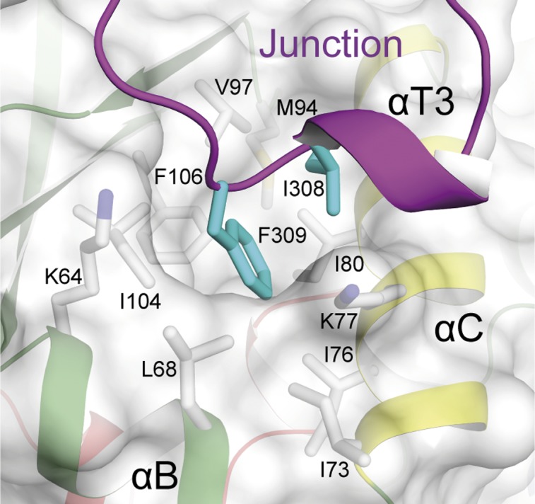
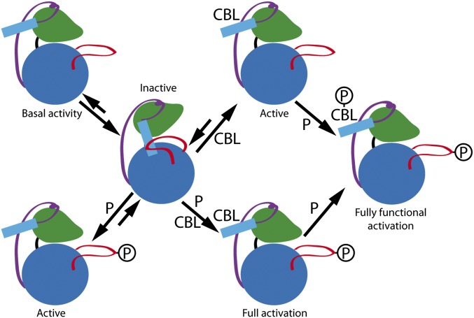
Plant cells have developed specific protective molecular machinery against environmental stresses. The family of CBL-interacting protein kinases (CIPK) and their interacting activators, the calcium sensors calcineurin B-like (CBLs), work together to decode calcium signals elicited by stress situations. The molecular basis of biological activation of CIPKs relies on the calcium-dependent interaction of a self-inhibitory NAF motif with a particular CBL, the phosphorylation of the activation loop by upstream kinases, and the subsequent phosphorylation of the CBL by the CIPK. We present the crystal structures of the NAF-truncated and pseudophosphorylated kinase domains of CIPK23 and CIPK24/SOS2. In addition, we provide biochemical data showing that although CIPK23 is intrinsically inactive and requires an external stimulation, CIPK24/SOS2 displays basal activity. This data correlates well with the observed conformation of the respective activation loops: Although the loop of CIPK23 is folded into a well-ordered structure that blocks the active site access to substrates, the loop of CIPK24/SOS2 protrudes out of the active site and allows catalysis. These structures together with biochemical and biophysical data show that CIPK kinase activity necessarily requires the coordinated releases of the activation loop from the active site and of the NAF motif from the nucleotide-binding site. Taken all together, we postulate the basis for a conserved calcium-dependent NAF-mediated regulation of CIPKs and a variable regulation by upstream kinases.

Keywords: abiotic stress; ion transport; signaling.


Conflict of interest statement

The authors declare no conflict of interest.

Figures

Fig. 1.
Fig. 1.
The crystal structures of CIPK23 and CIPK24/SOS2. (A) Domain structure of CIPKs. (B) A ribbon representation of the crystal structures of CIPK23ΔC T190D (Left) and CIPK24/SOS2ΔC T168D (Right). The key structural features are labeled and highlighted in different colors. (C) The ribbon representation of the active site section of the CIPK23ΔC T190D, CIPK24/SOS2ΔC T168D (semitransparent overlaid on the left), and the related active kinase PKA (Right) (PDB ID code: 1ATP). The catalytically relevant residues and the ATP are displayed in a stick representation; CIPK23 and CIPK24/SOS2 residues are displayed in white and cyan, respectively.
Fig. 2.
Fig. 2.
Comparative kinase activity analyses of CIPK23 and CIPK24/SOS2 proteins. The average specific activities expressed in nmol⋅min−1⋅μg−1 of protein units determined by a kinase spectrophotometric assay of the proteins expressed in E. coli. Error bars indicate the SD calculated from four independent measurements. All of the distributions are different compared with the CIPK23ΔC with a confidence level of 99% but those corresponding to CIPK23ΔC T190D and CIPK23-FILS in which the confidence level is 95% (Student’s t test).
Fig. 3.
Fig. 3.
The activation loop. (A) Sequence alignment of the activation loop of CIPKs together with a schematic representation of CIPK23ΔC T190D and CIPK24/SOS2ΔC T168D secondary structures. Sequences are grouped according to their sequence similarity. Residues are colored according to their conservation (77). The arrows highlight the totally conserved and potentially phosphorylable residues. The rectangular box highlights the residues at αT2 that serve to classify the CIPKs into four groups. (B) A detailed view of CIPK23ΔC T190D showing the structural environment of the conserved Ser176, Thr190, and Tyr197. The view highlights the role of Val182 and Arg183 in the stabilization of the loop at the active site. Inset shows a comparison of CIPK23ΔC (white sticks) and CIPK23ΔC T190D (cyan sticks) structures showing that the point mutation is accommodated without a significant change in the loop conformation.
Fig. 4.
Fig. 4.
(A) Ribbon and schematic representations of the CIPK24/SOS2 regulatory domain structures in complex with CBL4/SOS3 (PDB ID code: 2EHB) (Left) and CIPK23ΔC T190D together with a surface representation of the cavity connecting the ATP binding site and the hinge region between N and C lobes (Right). The cavity is wide enough to accommodate the two amphipathic helices forming the NAF motif. (B) Comparison of the far-UV CD spectra of CIPK23ΔC and CIPK23-NAF; the arrows indicate the characteristic α-helical maximum at 195, and minima at 208 and 222 nm. (C) Comparison of the thermal stability profiles monitored by CD for CIPK23ΔC (Left) and CIPK23-NAF (Right) at increasing CHAPS concentration.
Fig. 5.
Fig. 5.
A section of the CIPK23ΔC T190D structure showing the interaction between Ile308 and Phe309 with the hydrophobic pocket at the N lobe of CIPK23. A cartoon representation of the kinase is displayed together with a semitransparent surface.
Fig. 6.
Fig. 6.
A working model for CIPKs regulation. CIPK is schematically represented with the N lobe and the C lobe depicted in green and blue, respectively, the junction as a purple line, the activation loop in red, and the NAF motif as a cyan rectangle. Activation of CIPKs implies necessarily the release of both the activation loop and the self-inhibitory NAF motif. The calcium-dependent CBL interaction and/or phosphorylation of the activation loop set the equilibrium between the inactive and the active forms. The activity of CIPKs depends on the balance between active and inactive forms. Some CIPKs (for instance CIPK24/SOS2) display basal activity. The junction region may be responsible for the stabilization of a CBL unbound and unphosphorylated CIPK form with basal activity. Once CIPK is active, the fully functional activation is achieved by the phosphorylation of the CBL.

References

    1. Albrecht V, Ritz O, Linder S, Harter K, Kudla J. The NAF domain defines a novel protein-protein interaction module conserved in Ca2+-regulated kinases. EMBO J. 2001;20(5):1051–1063. - PMC - PubMed
    1. Shi J, et al. Novel protein kinases associated with calcineurin B-like calcium sensors in Arabidopsis. Plant Cell. 1999;11(12):2393–2405. - PMC - PubMed
    1. Batistič O, Kudla J. Analysis of calcium signaling pathways in plants. Biochim Biophys Acta. 2012;1820(8):1283–1293. - PubMed
    1. Steinhorst L, Kudla J. Calcium and reactive oxygen species rule the waves of signaling. Plant Physiol. 2013;163(2):471–485. - PMC - PubMed
    1. Luan S, Kudla J, Rodriguez-Concepcion M, Yalovsky S, Gruissem W. Calmodulins and calcineurin B-like proteins: Calcium sensors for specific signal response coupling in plants. Plant Cell. 2002;14(Suppl):S389–S400. - PMC - PubMed

Publication types

MeSH terms

LinkOut - more resources