. 2014 Oct;70(Pt 10):2667-74.
doi: 10.1107/S1399004714017064. Epub 2014 Sep 27.

Structures of human constitutive nitric oxide synthases

Affiliations

Structures of human constitutive nitric oxide synthases

Huiying Li et al. Acta Crystallogr D Biol Crystallogr. 2014 Oct.

Abstract

Mammals produce three isoforms of nitric oxide synthase (NOS): neuronal NOS (nNOS), inducible NOS (iNOS) and endothelial NOS (eNOS). The overproduction of NO by nNOS is associated with a number of neurodegenerative disorders; therefore, a desirable therapeutic goal is the design of drugs that target nNOS but not the other isoforms. Crystallography, coupled with computational approaches and medicinal chemistry, has played a critical role in developing highly selective nNOS inhibitors that exhibit exceptional neuroprotective properties. For historic reasons, crystallography has focused on rat nNOS and bovine eNOS because these were available in high quality; thus, their structures have been used in structure-activity-relationship studies. Although these constitutive NOSs share more than 90% sequence identity across mammalian species for each NOS isoform, inhibitor-binding studies revealed that subtle differences near the heme active site in the same NOS isoform across species still impact enzyme-inhibitor interactions. Therefore, structures of the human constitutive NOSs are indispensible. Here, the first structure of human neuronal NOS at 2.03 Å resolution is reported and a different crystal form of human endothelial NOS is reported at 1.73 Å resolution.

Keywords: nitric oxide synthase.


Figures

Figure 1
Figure 1
(a) Superimposition of bovine eNOS (purple) and human eNOS (yellow) structures. Heme (red), tetrahydrobiopterin (green) and l-Arg (blue) are shown as ball-and-stick representations. A few surface Gd and bis-tris clusters are highlighted in cyan. The overall r.m.s.d. for dimeric eNOS is 0.65 Å with 803 residues compared. (b) A surface Gd3+ site next to Glu321 in chain B stabilized by bis-tris. The 2F oF c density level for Gd3+ is higher than 40σ. The ligation bond distances from eight oxygen donors are marked in Å.
Figure 2
Figure 2
Two dimers in the asymmetric unit of human nNOS. Dimer AB packs tightly against dimer CD in a handshake manner.
Figure 3
Figure 3
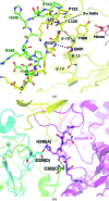
(a) Comparison of the surface loop in human nNOS (green) with that in human iNOS (yellow). The secondary structure of iNOS is also shown as a cartoon with a three-stranded β-sheet labeled (S-13, S-14, S-18). The backbone–backbone hydrogen bonds from the loop to an extension (residues 490 and 491) from the β-sheet are shown by dashed lines. In contract to the 310-helix (residues 122–125) observed in iNOS, the loop in nNOS is a random coil from Pro343 to Arg349. (b) The flexible loop in human nNOS is stabilized by some intersubunit packing contacts, which are shown as dashed lines.
Figure 4
Figure 4
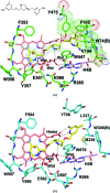
(a) The active site of human eNOS with an aminopyridine inhibitor bound. The OMIT F oF c density for the inhibitor is contoured at 3.0σ. Major hydrogen bonds are depicted as dashed lines. The hydrophobic pocket on the outside of the active site is lined with Val104, Phe105, Tyr475 and Trp74 from chain B and is highlighted with a surface dot representation. (b) The active site of rat nNOS with the same inhibitor bound (PDB entry 4c39; Jing et al., 2014 ▶) but in a double-headed binding mode with both aminopyridine rings involved in hydrogen bonds to protein and heme.
Figure 5
Figure 5
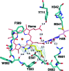
The active site of human nNOS with l-Arg bound. The 2F oF c electron density for l-Arg is contoured at 1.0σ. The extensive hydrogen-bonding networks involving protein, heme, H4B and l-Arg are depicted as dashed lines with distances marked in Å.
Figure 6
Figure 6
An aminoquinoline inhibitor bound to the rat nNOS active site (PDB entry 4cdt; Cinelli et al., 2014 ▶). For comparison, two residues from the human nNOS structure, Met341 and His342 (yellow), are overlaid. The potential clash between the fluorophenyl ring and the His342 side chain is obvious.

References

    1. Adams, P. D. et al. (2010). Acta Cryst. D66, 213–221. - PubMed
    1. Bredt, D. S., Ferris, C. D. & Snyder, S. H. (1992). J. Biol. Chem. 267, 10976–10981. - PubMed
    1. Cho, H. J., Xie, Q.-W., Calaycay, J., Mumford, R. A., Swiderek, K. M., Lee, T. D. & Nathan, C. (1992). J. Exp. Med. 176, 599–604. - PMC - PubMed
    1. Cinelli, M. A., Li, H., Chreifi, G., Martásek, P., Roman, L. J., Poulos, T. L. & Silverman, R. B. (2014). J. Med. Chem. 57, 1513–1530. - PMC - PubMed
    1. Crane, B. R., Arvai, A. S., Ghosh, D. K., Wu, C., Getzoff, E. D., Stuehr, D. J. & Tainer, J. A. (1998). Science, 279, 2121–2126. - PubMed

Publication types

MeSH terms