. 2012 Apr 24;51(16):3497-507.
doi: 10.1021/bi300307b. Epub 2012 Apr 9.

Structure and catalytic mechanism of LigI: insight into the amidohydrolase enzymes of cog3618 and lignin degradation

Affiliations

Structure and catalytic mechanism of LigI: insight into the amidohydrolase enzymes of cog3618 and lignin degradation

Merlin Eric Hobbs et al. Biochemistry. .

Abstract

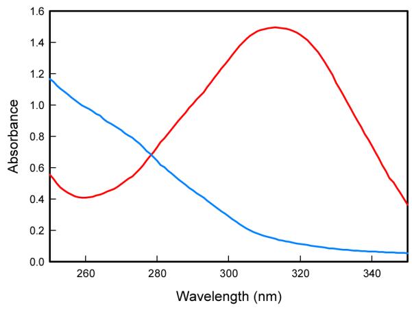
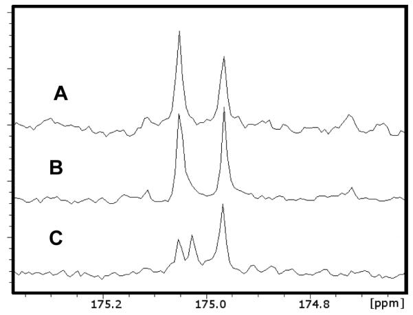
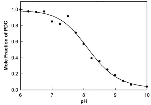
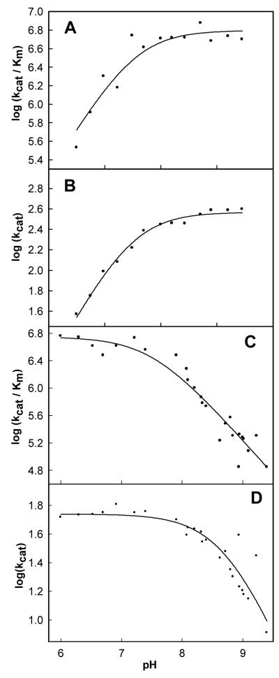
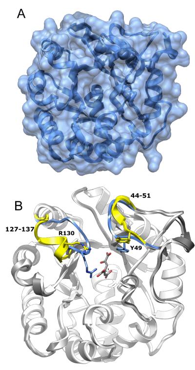
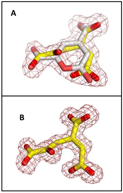
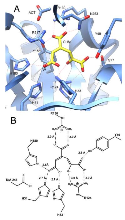
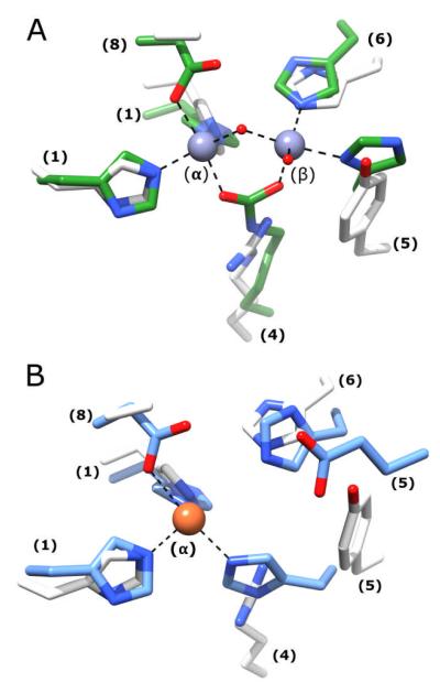
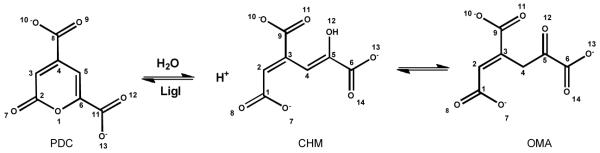
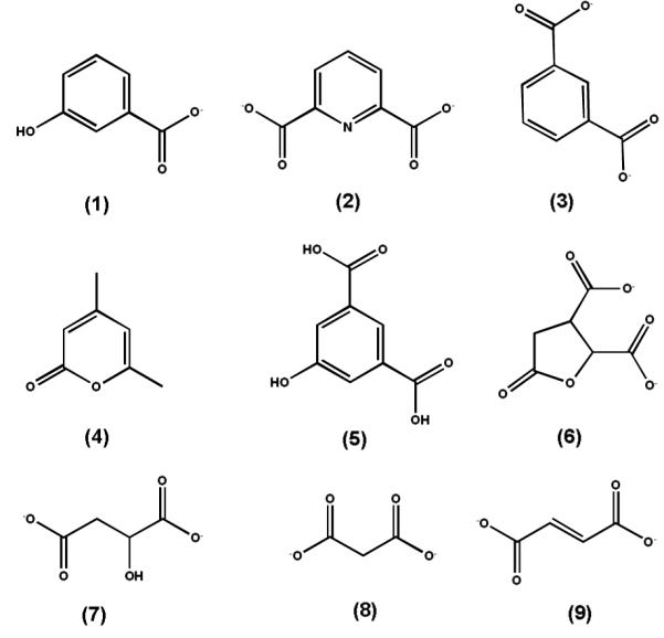
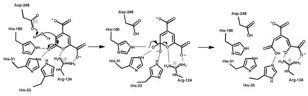
LigI from Sphingomonas paucimobilis catalyzes the reversible hydrolysis of 2-pyrone-4,6-dicarboxylate (PDC) to 4-oxalomesaconate and 4-carboxy-2-hydroxymuconate in the degradation of lignin. This protein is a member of the amidohydrolase superfamily of enzymes. The protein was expressed in Escherichia coli and then purified to homogeneity. The purified recombinant enzyme does not contain bound metal ions, and the addition of metal chelators or divalent metal ions to the assay mixtures does not affect the rate of product formation. This is the first enzyme from the amidohydrolase superfamily that does not require a divalent metal ion for catalytic activity. The kinetic constants for the hydrolysis of PDC are 340 s(-1) and 9.8 × 10(6) M(-1) s(-1) (k(cat) and k(cat)/K(m), respectively). The pH dependence on the kinetic constants suggests that a single active site residue must be deprotonated for the hydrolysis of PDC. The site of nucleophilic attack was determined by conducting the hydrolysis of PDC in (18)O-labeled water and subsequent (13)C nuclear magnetic resonance analysis. The crystal structures of wild-type LigI and the D248A mutant in the presence of the reaction product were determined to a resolution of 1.9 Å. The C-8 and C-11 carboxylate groups of PDC are coordinated within the active site via ion pair interactions with Arg-130 and Arg-124, respectively. The hydrolytic water molecule is activated by the transfer of a proton to Asp-248. The carbonyl group of the lactone substrate is activated by electrostatic interactions with His-180, His-31, and His-33.


Figures

Figure 1
Figure 1
Cytoscape generated sequence similarity network of cog3618 at a BLAST E-value cut-off of 10−70. Nodes represent proteins within cog3618 connected to other proteins with an E-value cutoff of less than 10−70 by lines. The stringency value of 10−70 was arbitrarily chosen based on what appears to be the presence of smaller isofunctional groups. The triangle shaped nodes colored red and blue in Group 3 repsresent LigI from S. paucimobilis and 4-SML from A. tumefaciens, respectively. Circular nodes colored green in Group 3 are annotated here as LigI, based on conservation of active site residues of LigI from S. paucimobilis. Nodes colored blue in Group 3 are predicted to be 4-SML enzymes. The yellow nodes in Group 3 are unannotated. The triangle shaped node colored blue in Group 8 is L-rhamnono-1,4-lactonase from Azotobacter vinelandii. The rest of the proteins are of unknown function.
Figure 2
Figure 2
UV-visible absorbance spectrum of 0.3 mM PDC (red) and the hydrolysis product after the addition of LigI (blue) at pH 10. The maximum absorbance change is at 312 nm with a differential extinction coefficient, Δε, of 6248 M−1 cm−1.
Figure 3
Figure 3
(A) NMR spectrum of OMA in water at pH 9.0. (B) NMR spectrum of OMA incubated in 50% 18O water at pH 9.0. (C) NMR spectrum of OMA produced enzymatically from PDC in 18O water at pH 9.0.
Figure 4
Figure 4
Effect of pH on the equilibrium concentrations of PDC and OMA/CHM. The line represents a fit of the data to equation 3. The equilibrium constant for the reaction illustrated in Scheme 1 is 5.7 × 10⁻⁹ M−1.
Figure 5
Figure 5
pH-rate profiles for the enzymatic hydrolysis and synthesis of PDC. (A) pH-rate profile for log (kcat/Km) for the hydrolysis of PDC. (B) pH-rate profile for log (kcat) for the hydrolysis of PDC. The lines for plots A and B represent the fit of the data to equation 4. (C) pH-rate profile for log (kcat/Km) for the synthesis of PDC. (D) pH-rate profile for log (kcat) for the synthesis of PDC. The lines for plots C and D represent the fit of the data to equation 5.
Figure 6
Figure 6
Structure of native LigI and the D248A mutant. (A) Native LigI. The narrow crevice on the top of the solvent accessible surface corresponds to the active site entrance. (B) View of CHM-bound D248A LigI (white) superimposed onto the wild-type apo-form of LigI (gray). The two loops, Phe-127 to Lys-137 and Ser-44 to Pro-51, that exhibit a change in conformation upon substrate binding, are color coded to the following format: Native LigI (apo) is yellow and CHM-bound D248A is blue.
Figure 7
Figure 7
Refined electron density (2FoFc, contoured at 1σ) of the ligand in the active site of LigI. (A) Presence of about equimolar amount of substrate (PDC, white) and product (CHM, yellow) at pH 8.5; (B) Presence of product only (CHM, yellow) at pH 6.5.
Figure 8
Figure 8
(A) CHM bound in the active site of the D248A mutant of LigI at pH 6.5. No PDC was detected at this pH. (B) Active site structure of D248A with CHM in the active site.
Figure 9
Figure 9
(A) The active site of D248A LigI is structurally aligned with the binuclear metal center of phosphotriesterase (PTE). The active site is color-coded to the following format: PTE, PDB code: 1hzy (green) and D248A LigI active site is shown in white. (B)The active site of D248A LigI is structurally aligned with the mononuclear metal center found in the active site of cytosine deaminase. The active sites are color-coded to the following format: CDA, PDB code: 1k70 (blue) and D248A LigI active site is shown in white. Numbers in parentheses indicate the β- strand origin of each residue.
Scheme 1
Scheme 1
Scheme 2
Scheme 2
Scheme 3
Scheme 3

References

    1. Hishida M, Shikinaka K, Katayama Y, Kajita S, Masai E, Nakamura M, Otsuka Y, Ohara S, Shigehara K. Polyesters of 2-pyrone-4,6-dicarboxylic acid (PDC) as bio-based plastics exhibiting strong adhering properties. Polymer Journal. 2009;41:297–302.
    1. Kersten PJ, Dagley S, Whittaker JW, Arciero DM, Lipscomb JD. 2-Pyrone-4,6-dicarboxylic acid, a catabolite of gallic acids in Pseudomonas species. Journal of Bacteriology. 1982;152:1154–1162. - PMC - PubMed
    1. Maruyama K. Purification and properties of 2-pyrone-4,6-dicarboxylate hydrolase. Journal of Biochemistry. 1983;93:557–565. - PubMed
    1. Holm L, Sander C. An Evolutionary Treasure: Unification of a Broad Set of Amidohydrolases Related to Urease. Proteins: Struct., Funct., Genet. 1997;28:72–82. - PubMed
    1. Seibert CM, Raushel FM. Structural and Catalytic Diversity within the Amidohydrolase Superfamily. Biochemistry. 2005;44:6383–6391. - PubMed

MeSH terms

LinkOut - more resources