. 2012 Oct 10;20(10):1788-95.
doi: 10.1016/j.str.2012.08.011. Epub 2012 Sep 6.

The CDK9 tail determines the reaction pathway of positive transcription elongation factor b

Affiliations

The CDK9 tail determines the reaction pathway of positive transcription elongation factor b

Sonja Baumli et al. Structure. .

Abstract

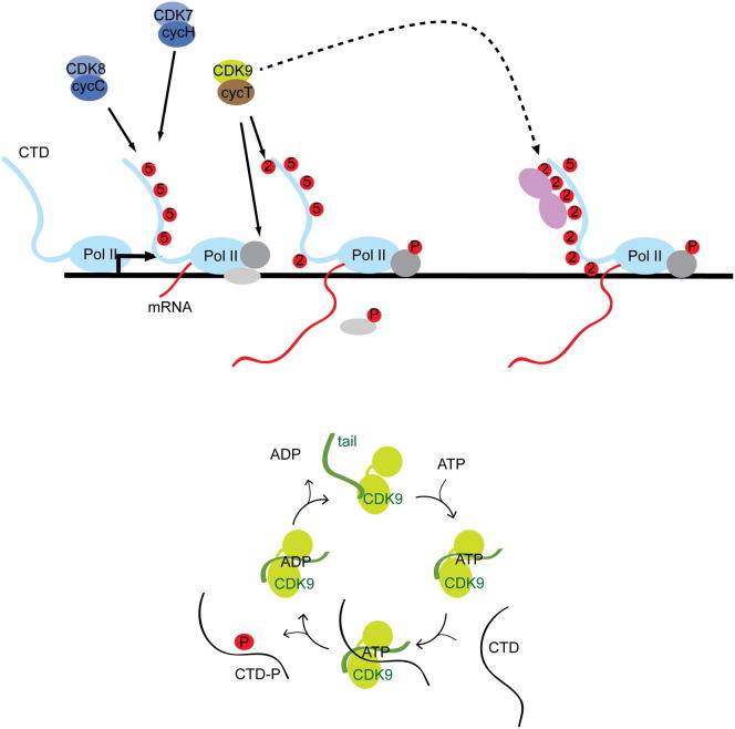
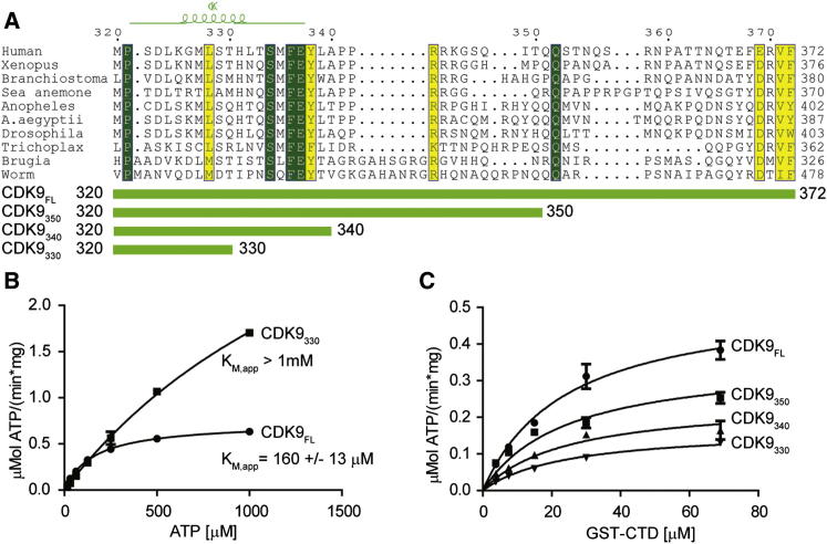
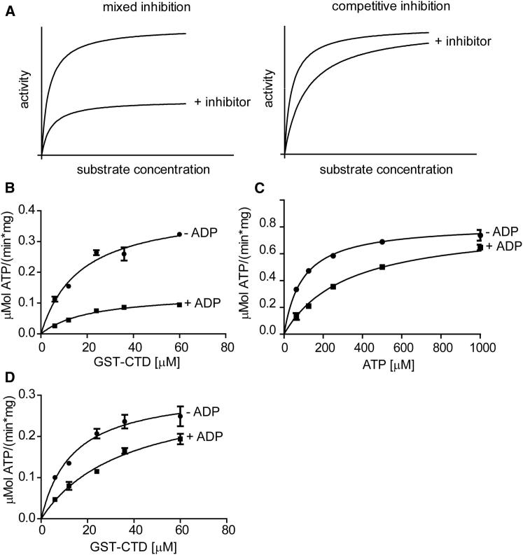
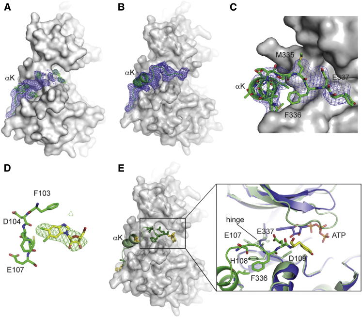
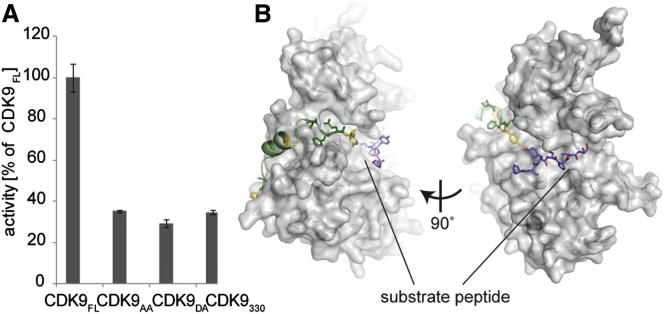
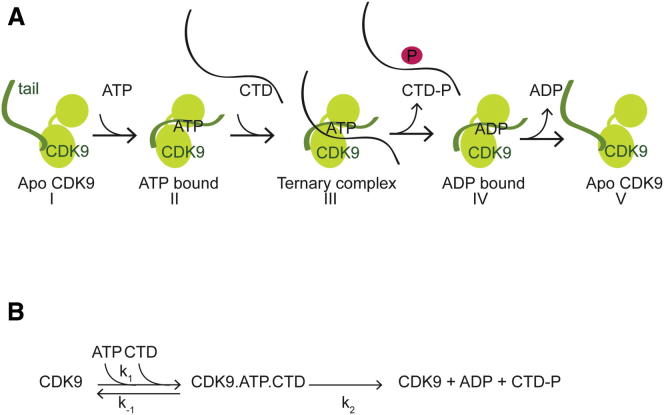
CDK9, the kinase of positive transcription elongation factor b (P-TEFb), stimulates transcription elongation by phosphorylating RNA polymerase II and transcription elongation factors. Using kinetic analysis of a human P-TEFb complex consisting of CDK9 and cyclin T, we show that the CDK9 C-terminal tail sequence is important for the catalytic mechanism and imposes an ordered binding of substrates and release of products. Crystallographic analysis of a CDK9/cyclin T complex in which the C-terminal tail partially blocks the ATP binding site reveals a possible reaction intermediate. Biochemical characterization of CDK9 mutants supports a model in which the CDK9 tail cycles through different conformational states. We propose that this mechanism is critical for the pattern of CTD Ser2 phosphorylation on actively transcribed genes.


Figures

None
Graphical abstract
Figure 1
Figure 1
The C Terminus of CDK9 Influences Kinase Activity (A) Sequence alignment of the C-terminal region of CDK9. Identical residues are highlighted in green, and chemically equivalent residues are in yellow. The location of the most C-terminal CDK9 α helix is shown above the alignment. The constructs used in (B) and (C) are identified below the alignment. (B) Activity of CDK9FL/cyclin T and CDK9330/cyclin T assayed in the presence of increasing amounts of ATP. (C) Activity of various CDK9/cyclin T complexes with increasing concentrations of GST-CTD. CDK9 variants are labeled as shown in (A). All measurements were done in triplicate and reproduced in independent experiments. Error bars indicate standard errors (SEs). For a summary of the apparent kinetic parameters, see Table S1. See also Figure S1.
Figure 2
Figure 2
The CDK9 Tail Is Required for the Ordered Substrate Addition Catalytic Mechanism (A) Theoretical model curves for mixed and competitive inhibition assuming the same KM, Ki, and Vmax in both cases. (B) Activity of CDK9FL/cyclin T in the absence and presence of 2.5 μM ADP, in the presence of 100 μM ATP and increasing amounts of CTD. (C) Activity of CDK9FL/cyclin T in the absence and presence of 2.5 μM ADP, in the presence of 36 μM CTD and increasing amounts of ATP. (D) Activity of CDK9330/cyclin T in the absence and presence of 2.5 μM ADP, in the presence of 100 μM ATP and increasing amounts of CTD. All measurements were done in triplicate and reproduced in independent experiments. Error bars in (B)–(D) represent SEs. See also Figure S3.
Figure 3
Figure 3
Structure of the CDK9 C-Terminal Tail (A) The final electron density map corresponding to additional C-terminal residues present in CDK9FL bound to cyclin T259 in the absence of DRB or AMPPNP is shown as a blue mesh at a contour level of 1σ. The difference density is shown as a green mesh at a contour level of 3σ. The CDK9 core structure is displayed as a solvent-accessible surface representation in gray. (B) Same view as in (A) to show the electron density corresponding to the CDK9 C-terminal sequence present in the structure of CDK9FL/cyclin T259 bound to DRB. The electron density map is drawn as a blue mesh and contoured at 1σ. (C) Detailed view of the interactions between the CDK9 C-terminal residues and the hinge sequence present in the CDK9FL/cyclin T259/DRB complex. The solvent-accessible surface of CDK9330 is drawn in gray and selected residues are labeled. The electron density map of the refined structure is drawn at a contour level of 1σ. (D) The difference density map corresponding to the DRB binding site in CDK9FL is shown at a contour level of 3σ. Residues of the CDK9FL hinge region (from Phe103 to Glu107) are also shown. (E) Interactions of Phe336 and Glu337 with the CDK9 hinge region. C-terminal residues that are identical across species are drawn as sticks in green, and equivalent residues are in yellow. CDK9/cyclin T259 (PDB 3BLH) bound to ATP is superposed in blue for comparison. See also Figure S4.
Figure 4
Figure 4
Flexibility of the CDK9 C Terminus Is Important for Kinase Activity (A) Activity of CDK9FL/cyclin T toward GST-CTD compared with complexes containing the CDK9 clamp mutants CDK9AA and CDK9DA, and the CDK9 truncation CDK9330. All measurements were done at 100 μM ATP and 24 μM GST-CTD in triplicate and reproduced in independent experiments. Error bars represent SEs. (B) Model showing that binding of a substrate peptide (lilac) is compatible with the bound conformation of the CDK9 C-terminal region. In this model, Ser2 of the heptad repeat occupies the phosphotransfer position. The cartoon-and-stick model is as described in Figure 3E.
Figure 5
Figure 5
Model of the CDK9 Catalytic Cycle (A) Model of the CDK9 conformational states adopted during the catalytic cycle. The cyclin subunit is omitted from the scheme for clarity. (I) Apo CDK9 has a flexible C-terminal tail. ATP is the first substrate bound to CDK9. (II) Upon ATP binding, the CDK9 C-terminal tail adopts an ordered structure, partially covering the ATP binding site. (III) The Pol II CTD binds to the CDK9/ATP complex to form a ternary complex that is competent for phosphotransfer. (IV) After phosphorylation, the CTD substrate is released. (V) ADP is released and the kinase returns to its apo state with a flexible C-terminal tail. (B) Reaction scheme. k1 and k-1 refer to the rate constants for ternary complex formation, and k2 is the rate constant governed by phosphotransfer and product release.

References

    1. Adams J.A. Kinetic and catalytic mechanisms of protein kinases. Chem. Rev. 2001;101:2271–2290. - PubMed
    1. Adams P.D., Afonine P.V., Bunkóczi G., Chen V.B., Davis I.W., Echols N., Headd J.J., Hung L.W., Kapral G.J., Grosse-Kunstleve R.W. PHENIX: a comprehensive Python-based system for macromolecular structure solution. Acta Crystallogr. D Biol. Crystallogr. 2010;66:213–221. - PMC - PubMed
    1. Anand K., Schulte A., Fujinaga K., Scheffzek K., Geyer M. Cyclin box structure of the P-TEFb subunit cyclin T1 derived from a fusion complex with EIAV tat. J. Mol. Biol. 2007;370:826–836. - PMC - PubMed
    1. Bartkowiak B., Liu P., Phatnani H.P., Fuda N.J., Cooper J.J., Price D.H., Adelman K., Lis J.T., Greenleaf A.L. CDK12 is a transcription elongation-associated CTD kinase, the metazoan ortholog of yeast Ctk1. Genes Dev. 2010;24:2303–2316. - PMC - PubMed
    1. Baumli S., Lolli G., Lowe E.D., Troiani S., Rusconi L., Bullock A.N., Debreczeni J.E., Knapp S., Johnson L.N. The structure of P-TEFb (CDK9/cyclin T1), its complex with flavopiridol and regulation by phosphorylation. EMBO J. 2008;27:1907–1918. - PMC - PubMed

Publication types

MeSH terms