. 2014 Nov 24;207(4):481-98.
doi: 10.1083/jcb.201408111. Epub 2014 Nov 17.

A network of assembly factors is involved in remodeling rRNA elements during preribosome maturation

Affiliations

A network of assembly factors is involved in remodeling rRNA elements during preribosome maturation

Jochen Baßler et al. J Cell Biol. .

Erratum in

Abstract

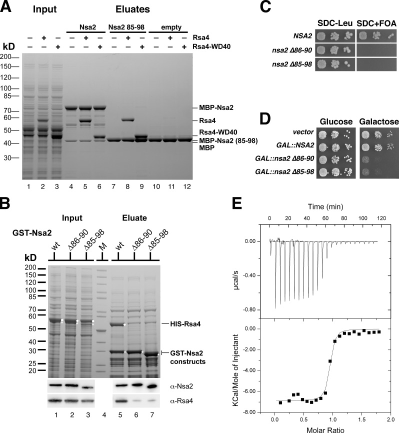
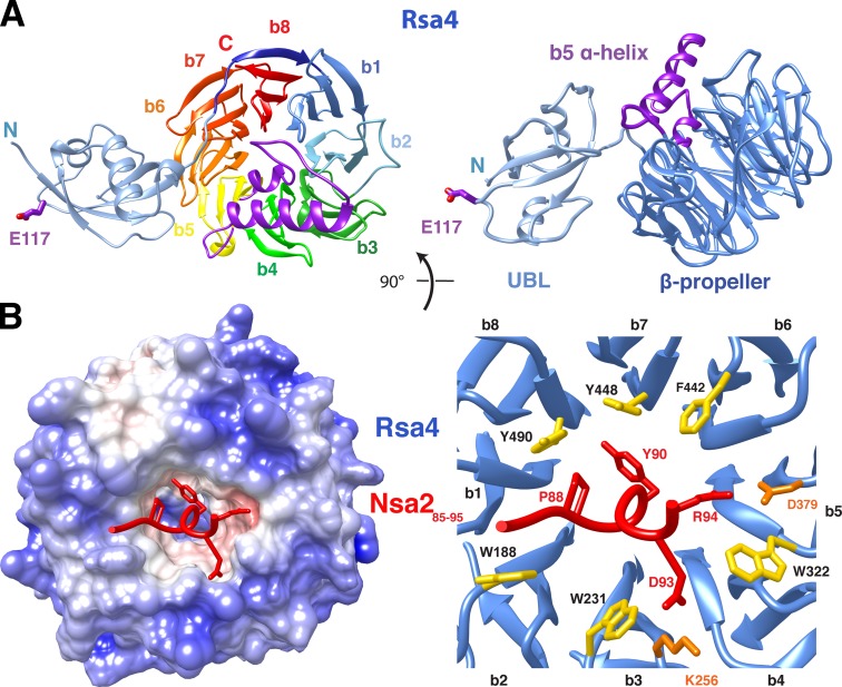
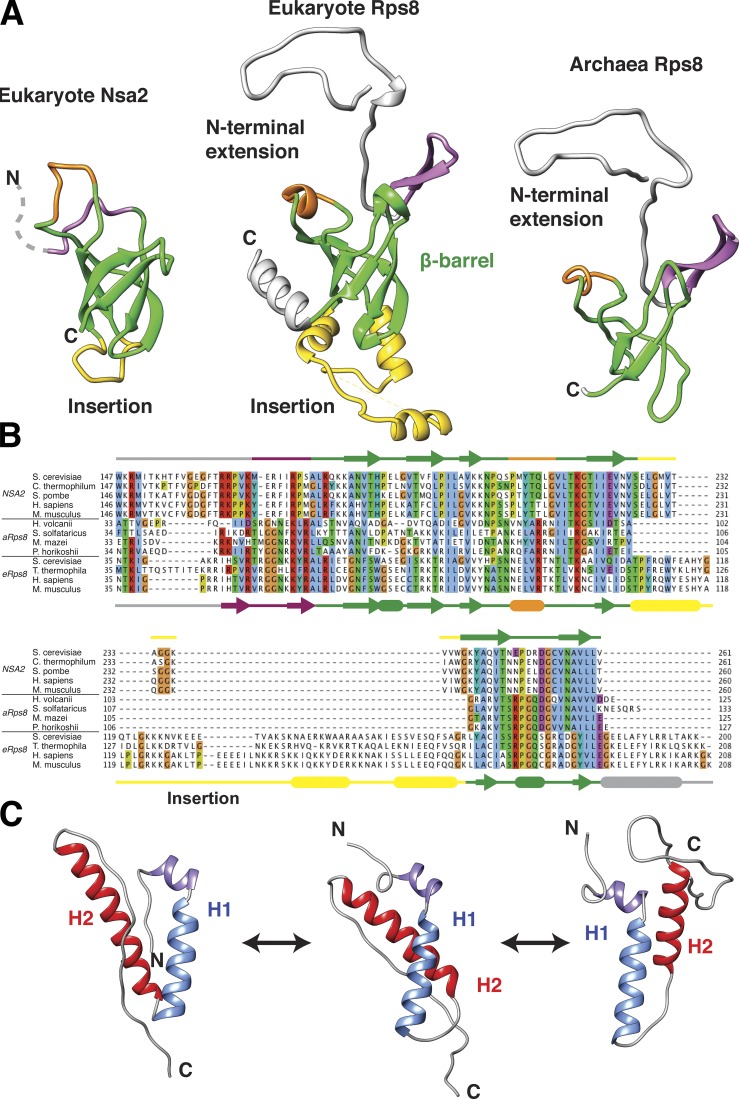
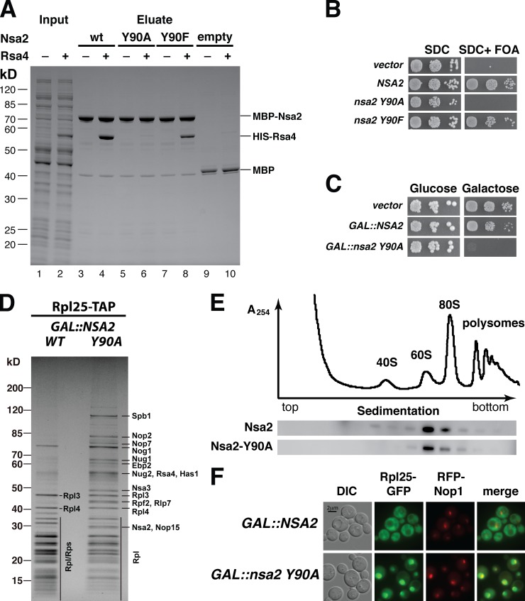
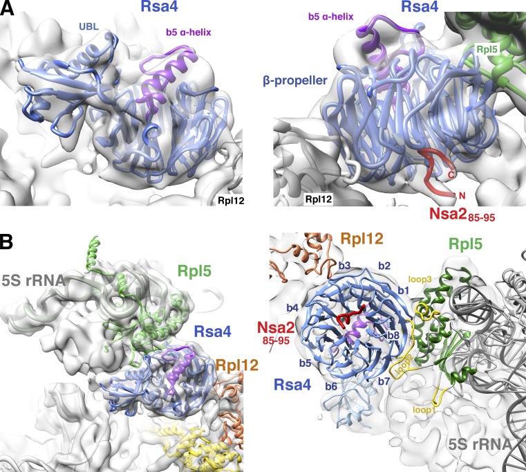
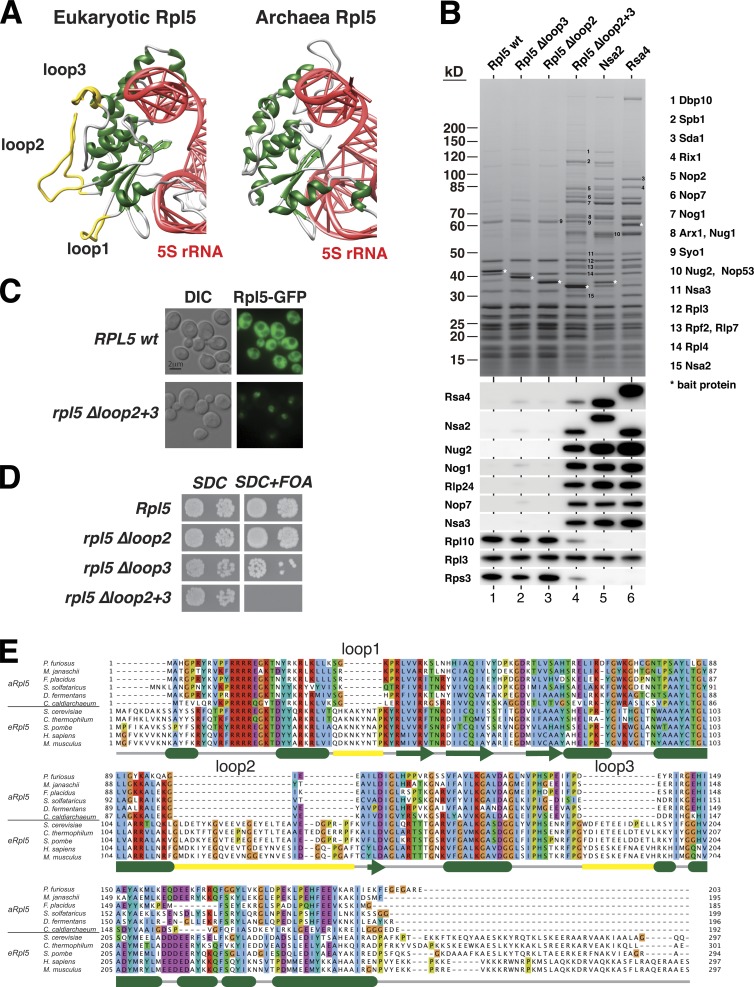
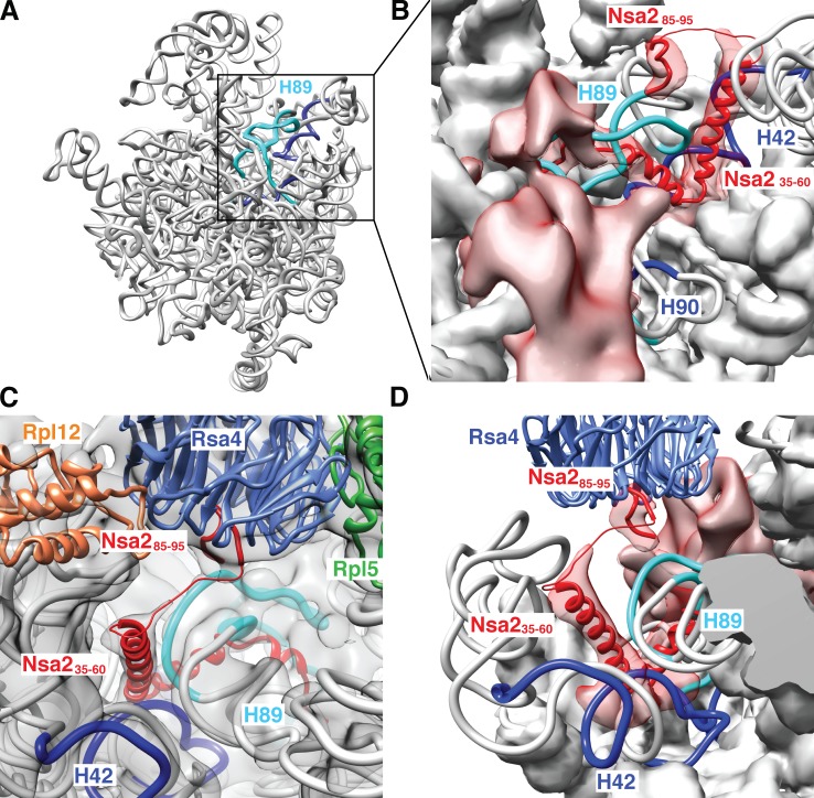
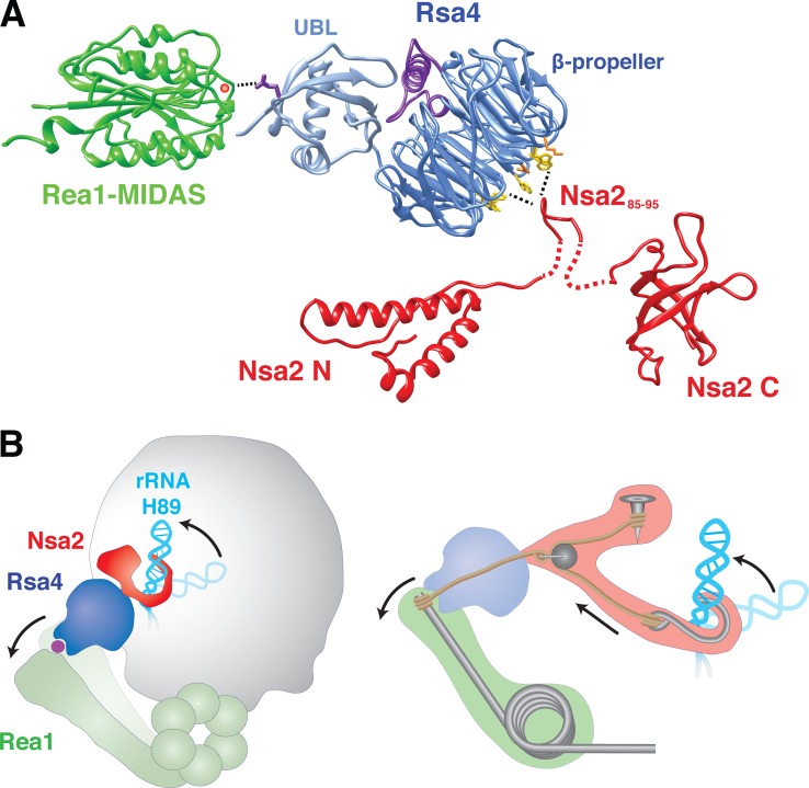
Eukaryotic ribosome biogenesis involves ∼200 assembly factors, but how these contribute to ribosome maturation is poorly understood. Here, we identify a network of factors on the nascent 60S subunit that actively remodels preribosome structure. At its hub is Rsa4, a direct substrate of the force-generating ATPase Rea1. We show that Rsa4 is connected to the central protuberance by binding to Rpl5 and to ribosomal RNA (rRNA) helix 89 of the nascent peptidyl transferase center (PTC) through Nsa2. Importantly, Nsa2 binds to helix 89 before relocation of helix 89 to the PTC. Structure-based mutations of these factors reveal the functional importance of their interactions for ribosome assembly. Thus, Rsa4 is held tightly in the preribosome and can serve as a "distribution box," transmitting remodeling energy from Rea1 into the developing ribosome. We suggest that a relay-like factor network coupled to a mechano-enzyme is strategically positioned to relocate rRNA elements during ribosome maturation.


Figures

Figure 1.
Figure 1.
A short peptide within Nsa2 is essential for binding to Rsa4. (A) In vitro binding assay of MBP-Nsa2 with Rsa4. Purified MBP-Nsa2 (lane 4–6), MBP-Nsa2 84–96 aa (lane 7–9), and MBP (lane 10–12) were immobilized on amylose beads and incubated with E. coli lysate without or with HIS6-TEV-Rsa4 full-length or HIS6-Rsa4 WD40 domain, respectively (lane 1–3). Eluates were analyzed by 4–12% SDS-PAGE and stained with Coomassie blue. (B) In vitro copurification of GST-Nsa2 and Rsa4. GST-Nsa2 wild-type, GST-Nsa2Δ86–90, or GST-Nsa2Δ85–98 were coexpressed with HIS-Rsa4 (input; see lanes 1, 2, and 3), purified via GSH Sepharose, and eluted by TEV cleavage (lanes 5, 6, and 7). The 4–12% SDS-polyacrylamide gel was stained with Coomassie blue, and Rsa4 and Nsa2 were detected by Western blot analysis. (C) Viability of nsa2 deletion mutants. An Nsa2 shuffle strain was transformed with plasmids encoding the indicated NSA2 alleles tagged with FTpA. Transformants were analyzed for complementation by spotting a 1:10 dilution series on SDC+FOA. The growth phenotype after 3 d of incubation at 30°C is shown. (D) Dominant-negative phenotype of nsa2 deletion mutants. Wild-type strain W303 was transformed with plasmids expressing the indicated NSA2 alleles tagged with FTpA under the control of the inducible GAL10 promoter. The toxic effect of NSA2 overexpression was tested on galactose-containing medium (SGC-TRP) after incubation for 3 d at 30°C. (E) ITC measurement of Rsa4 β-propeller with Nsa2 peptide is shown. Recombinant Rsa4 β-propeller (Rsa4Δ136) was expressed in E. coli, affinity-purified, and further purified by SEC before ITC measurement was performed with synthesized Nsa2 peptide (85–95 aa, DALPTYLLDRE).
Figure 2.
Figure 2.
Structural insight into the Rsa4–Nsa2 interaction. (A) Crystal structure of ctRsa4. Rsa4 consists of a UBL-like domain (light blue) followed by an eight-bladed β-propeller (left, b1–b8 in rainbow colors from dark blue to red; right, β-propeller rotated by 90° in dark blue) harboring the indicated α-helical insertion (purple) within blade 5. The highly conserved E117 (E114 in yeast) is also depicted, exposed on the surface of the UBL domain. (B) Crystal structure of the minimal scRsa4–scNsa2 complex. The surface view of scRsa4 is colored according to the charge calculated by PDB2PQR and APBS implemented in UCSF Chimera (left) with scNsa2 peptide (residues 86–95, red) bound into a hydrophobic pocket at the top site of the scRsa4 β-propeller (left). Also shown is a ribbon representation of the Nsa2 peptide (red) with its protruding residues bound to the scRsa4 β-propeller (blue) with hydrophobic (yellow) and charged residues involved in polar interactions (orange) at the rim of the propeller (right).
Figure 3.
Figure 3.
NMR solution structures of ctNsa2 domains. (A) NMR structure of recombinant ctNsa2-C (168–261 aa) adopts an Rps8 (eS8)-like six-stranded β-barrel fold. A structural similarity search of the protein databank performed with the DALI server identified the 40S ribosomal protein Rps8 (PDB accession no. 3U5C, chain I) from S. cerevisiae (S288c; sequence identity of 22%, Z score = 5.3, RMSD 3.7 Å) as structurally similar to ctNsa2 168–261 aa. The right panel shows the structure of an archaeal Rps8 taken from the PDB (accession no. 3J43). The six-stranded β-barrel fold is shown in green; variable insertions are shown in yellow, orange, and purple. (B) Multiple sequence alignment including Nsa2, archaeal Rps8 (aRps8), and eukaryotic Rps8 (eRps8). Secondary structure elements are indicated for ctNsa2 on top (derived by NMR) and S. cerevisiae Rps8 (PDB accession no. 3U5C) below. (C) Representative NMR structures of ctNsa2-N, determined by CS-Rosetta, show a common fold composed of one short N-terminal α-helix followed by two longer α-helices (H1 and H2). Multiple structures that differ in the packing and orientation of helix 2 reflect the disorder in the linker connecting the helices and the dynamic nature of Nsa2-N in solution.
Figure 4.
Figure 4.
The Rsa4–Nsa2 interaction is essential for ribosome biogenesis. (A) In vitro reconstitution of the Nsa2–Rsa4 interaction. Purified MBP-Nsa2 wild-type (wt; lanes 3 and 4), Nsa2 Y90A (lanes 5 and 6), Nsa2 Y90F (lanes 7 and 8), and MBP alone (lanes 9 and 10) were immobilized on amylose beads and incubated with E. coli lysate with or without HIS-TEV-Rsa4 (for input, see lanes 1 and 2). Eluates were analyzed by 4–12% SDS-PAGE and Coomassie staining, and the positions of the bands are indicated. (B) The highly conserved tyrosine 90 in Nsa2 is essential for yeast cell growth. Nsa2 shuffle strain was transformed with plasmids carrying the NSA2 or the indicated nsa2 mutant alleles. Complementation was analyzed by incubation of transformants on SDC+FOA plates at 30°C for 2 d. (C) Overexpression of nsa2 Y90A is dominant lethal. A yeast wild-type strain with endogenous NSA2 was transformed with 2μ plasmids carrying NSA2 or nsa2 Y90A alleles under the control of the galactose-inducible GAL promoter. The dominant-negative phenotype by NSA2 overexpression was tested on galactose-containing plates after incubation for 3 d at 30°C. (D) Dominant-lethal nsa2 Y90A induction arrests 60S biogenesis. Pulse-chase analysis of HA-Rpl25-Flag-ProtA (uL23) isolated from cells expressing dominant-negative nsa2 Y90A and wild-type Nsa2. HA-Rpl25-Flag-ProtA was pulsed for 7 min with Ome-Tyr after a 60-min galactose induction, and subsequently chased for 20 min with glucose/tetracycline in cells expressing GAL::NSA2 (WT) or GAL::nsa2 Y90A (Y90A). Affinity-purified HA-Rpl25-Flag-ProtA was analyzed by SDS-PAGE and Coomassie staining, and bands were identified by mass spectrometry. (E) Mutant Nsa2 Y90A is efficiently assembled into preribosomes. Whole-cell lysates derived from wild-type cells expressing plasmid-borne NSA2-FTpA and nsa2 Y90A-FTpA, respectively, were fractionated on a 10–50% sucrose gradient, and fractions were analyzed by Western blotting using anti-ProtA antibodies to detect wild-type and mutant Nsa2 proteins. (F) Dominant-lethal nsa2 Y90A induction causes a defect in 60S subunit export. Wild-type yeast cells were transformed with plasmids harboring the 60S reporter Rpl25-EGFP (uL23) and mRFP-Nop1, and 2μ plasmids harboring GAL::NSA2 or GAL::nsa2 Y90A alleles, respectively. Cells were shifted to galactose-containing medium for 6 h before intracellular localization of Rpl25-EGFP, and mRFP-Nop1 was analyzed by fluorescence microscopy.
Figure 5.
Figure 5.
Contact of Rsa4 to Nsa2, Rpl5 (uL18), and Rpl12 (uL11) on the pre-60S subunit. The crystal structures of Rsa4 (blue) and the Nsa2 peptide (residues 85–96; red) were fit into the 8.7-Å resolution cryo-EM structure of the Arx1 pre-60S particle. (A) Close view of the electron density of Rsa4 from the subunit joining (left) and solvent site (right). (B) Contact of Rsa4 to neighboring proteins Rpl5 (uL18, green) and Rpl12 (uL11, orange) viewed from the subunit joining site (left) and from the opposite site (right).
Figure 6.
Figure 6.
The eukaryote-specific loops of Rpl5 (uL18) are essential for ribosome biogenesis. (A) The structure of eukaryotic Rpl5 (uL18, PDB accession no. 3U5I) from S. cerevisiae in comparison with archaeal Rpl5 (PDB 3J44). Eukaryote-specific loops are highlighted in yellow; 5S rRNA is shown in red. (B) Affinity purification of Rpl5 wild-type and loop mutant constructs in comparison to other purified bait proteins co-enriched with pre-60S particles. The indicated Flag-TEV-ProtA (FTpA) Rpl5 proteins were purified from yeast (lanes 1–4) and compared with distinct pre-60S particles affinity-purified via Nsa2-FTpA and FTpA-Rsa4 (lanes 5 and 6). The top panel shows a 4–12% SDS-polyacrylamide gel stained with Coomassie blue, with labeled proteins identified by mass spectroscopy. The bottom panel shows a Western blot analysis of the gel using the indicated antibodies. (C) In vivo localization of Rpl5 Δloop2+3. A wild-type strain was transformed with a plasmid expressing Rpl5-GFP or Rpl5 Δloop2+3-GFP. Fluorescence microcopy was performed to determine their localization. (D) Viability of rpl5 loop mutants. A RPL5 shuffle strain was transformed with the indicated rpl5 alleles. Complementation analysis was done by plating transformants onto SDC+FOA plates. Growth on SDC-LEU plates (SDC) is shown after 3 d, whereas growth on FOA plates is shown after a 5-d incubation at 30°C. (E) Multiple sequence alignment of Rpl5 (uL18) derived from archaea (aRpl5) and eukaryotes (eRpl5). Secondary structure elements of Rpl5 from S. cerevisiae are indicated with the same color code as in A.
Figure 7.
Figure 7.
Nsa2 contacts immature rRNA helix 89 in the pre-60S particle. (A) Nsa2 cross-linked rRNA fragments are shown in a 3D model of the 25S pre-rRNA (see also Fig. S5). The identified rRNA cross-link sites are highlighted (H89 in light blue; H42 and H90 in dark blue) as an rRNA ribbon present in the Arx1 pre-60S particle. (B and D) Enlarged view of the cross-linked region of the pre-60S particle with fits of the indicated sequences into assigned (Nsa2 residues 85–96; Nsa2 residues 35–60) and unassigned electron densities (volumes colored in red). rRNA is shown as artificial density (8 Å resolution) except for cross-linked H42, H89, and H90, which are depicted as a ribbon model. B is a view from the subunit-joining side, and C and D are from the solvent side. C shows the complete electron density including Rpl12 (uL11, orange), Rpl5 (uL18, green), and Rsa4 (blue) with bound Nsa2 (85–96) peptide (red). From here, unassigned Nsa2 residues (thin red line) connect to a nearby electron density in which Nsa235–60 α-helix is fitted (red). An overview of the complete pre-60S particle including neighbors of the Rsa4–Nsa2 pair is shown in Fig. S5 C.
Figure 8.
Figure 8.
Model of the factor relay between Nsa2, Rsa4, and the Rea1 MIDAS and its proposed function in rearranging helix 89 toward the PTC. (A) The depicted Rea1 MIDAS domain (green) carrying a coordinated cation (red ball) is a structural model (modeled with Phyre2, template PDB accession no. 4FX5). The ctRsa4 crystal structure is shown in blue, with conserved E117 orientated toward the MIDAS domain/ion. The Nsa2 N and C domains solved by NMR are shown in red (see also Fig. 3), and the Nsa2 peptide (85–95 aa) in proximity to the top site of the Rsa4 β-propeller with hydrophobic residues is shown in yellow. (B) Illustration of the proposed force relay that transmits mechano-chemical energy from Rea1 through Rsa4 and Nsa2 to helix 89.

References

    1. Adams P.D., Afonine P.V., Bunkóczi G., Chen V.B., Davis I.W., Echols N., Headd J.J., Hung L.W., Kapral G.J., Grosse-Kunstleve R.W., et al. . 2010. PHENIX: a comprehensive Python-based system for macromolecular structure solution. Acta Crystallogr. D Biol. Crystallogr. 66:213–221 10.1107/S0907444909052925 - DOI - PMC - PubMed
    1. Bassler J., Kallas M., and Hurt E.. 2006. The NUG1 GTPase reveals an N-terminal RNA-binding domain that is essential for association with 60 S pre-ribosomal particles. J. Biol. Chem. 281:24737–24744 10.1074/jbc.M604261200 - DOI - PubMed
    1. Baßler J., Grandi P., Gadal O., Lessmann T., Petfalski E., Tollervey D., Lechner J., and Hurt E.. 2001. Identification of a 60S preribosomal particle that is closely linked to nuclear export. Mol. Cell. 8:517–529 10.1016/S1097-2765(01)00342-2 - DOI - PubMed
    1. Baßler J., Kallas M., Pertschy B., Ulbrich C., Thoms M., and Hurt E.. 2010. The AAA-ATPase Rea1 drives removal of biogenesis factors during multiple stages of 60S ribosome assembly. Mol. Cell. 38:712–721 10.1016/j.molcel.2010.05.024 - DOI - PMC - PubMed
    1. Ben-Shem A., Garreau de Loubresse N., Melnikov S., Jenner L., Yusupova G., and Yusupov M.. 2011. The structure of the eukaryotic ribosome at 3.0 Å resolution. Science. 334:1524–1529 10.1126/science.1212642 - DOI - PubMed

Publication types

MeSH terms

Substances