. 2014 Oct;58(10):6122-32.
doi: 10.1128/AAC.02574-13. Epub 2014 Aug 4.

Structure, activity, and inhibition of the Carboxyltransferase β-subunit of acetyl coenzyme A carboxylase (AccD6) from Mycobacterium tuberculosis

Affiliations

Structure, activity, and inhibition of the Carboxyltransferase β-subunit of acetyl coenzyme A carboxylase (AccD6) from Mycobacterium tuberculosis

Manchi C M Reddy et al. Antimicrob Agents Chemother. 2014 Oct.

Abstract

In Mycobacterium tuberculosis, the carboxylation of acetyl coenzyme A (acetyl-CoA) to produce malonyl-CoA, a building block in long-chain fatty acid biosynthesis, is catalyzed by two enzymes working sequentially: a biotin carboxylase (AccA) and a carboxyltransferase (AccD). While the exact roles of the three different biotin carboxylases (AccA1 to -3) and the six carboxyltransferases (AccD1 to -6) in M. tuberculosis are still not clear, AccD6 in complex with AccA3 can synthesize malonyl-CoA from acetyl-CoA. A series of 10 herbicides that target plant acetyl-CoA carboxylases (ACC) were tested for inhibition of AccD6 and for whole-cell activity against M. tuberculosis. From the tested herbicides, haloxyfop, an arylophenoxypropionate, showed in vitro inhibition of M. tuberculosis AccD6, with a 50% inhibitory concentration (IC50) of 21.4 ± 1 μM. Here, we report the crystal structures of M. tuberculosis AccD6 in the apo form (3.0 Å) and in complex with haloxyfop-R (2.3 Å). The structure of M. tuberculosis AccD6 in complex with haloxyfop-R shows two molecules of the inhibitor bound on each AccD6 subunit. These results indicate the potential for developing novel therapeutics for tuberculosis based on herbicides with low human toxicity.


Figures

FIG 1
FIG 1
The reaction catalyzed by ACC proceeds in two steps. The biotin carboxylase (BC) step involves the carboxylation of a biotin molecule bound to a biotin carboxylase carrier protein (BCCP), followed by the transfer of the carboxyl moiety to an acetyl-CoA molecule, catalyzed by a carboxyltransferase (CT; M. tuberculosis AccD6), forming the product malonyl-CoA.
FIG 2
FIG 2
M. tuberculosis AccD6 in vitro assays. (A) Reaction velocity plotted versus malonyl-CoA concentration (Km = 390 ± 70 μM; Vmax = 5.5 ± 0.4 μM min−1). (B) IC50 (70.2 ± 1 μM) plot of racemic haloxyfop inhibition of M. tuberculosis AccD6. (C) IC50 (21.4 ± 1 μM) plot of haloxyfop-R inhibition of M. tuberculosis AccD6.
FIG 3
FIG 3
Ribbon diagram of M. tuberculosis AccD6. (A) Ribbon diagram of the two subunit AccD6 holoenzyme, with the haloxyfop ligands depicted as sticks and balls. Subunits are depicted by differences in color, both colored by secondary structure. Subunit 1 is in blue and subunit 2 is yellow. (B) Ribbon diagram (colored by secondary structure) of the apo AccD6 single subunit. (C) Superimposition of M. tuberculosis AccD6 subunit 1 (blue) and subunit 2 (tan). Cα RMSD, 1.0 Å. Subunits are shown as ribbons.
FIG 4
FIG 4
M. tuberculosis AccD6 active site. (A) Two sets of oxyanion-stabilizing residues are highly conserved among the CT domains of different species, including the NH of Gly137 and Gly138 and the NH of Gly336′ and Ala367′. The structural similarity strongly suggests a similar enzyme mechanism. (B) Comparison of the M. tuberculosis AccD6 and yeast ACC (PDB code 1OD2) active sites. The yeast structure is in blue, while the M. tuberculosis structure is in tan and biotin is in tan. Side chains within 4 Å of each ligand are depicted as sticks.
FIG 5
FIG 5
(A) Electron density of haloxyfop (yellow sticks) and composite OMIT map electron density (blue contoured at 1 σ). (B) Haloxyfop interaction with M. tuberculosis AccD6 binding site 1; (C) haloxyfop interaction with M. tuberculosis AccD6 at binding site 2. Haloxyfop is in yellow, and protein side chains are in green. Dashed lines represent hydrogen bonds, and numbers represent distances, in Å.
FIG 6
FIG 6
Haloxyfop interactions with residues on binding site 1 (A) and binding site 2 (B). Hydrogen bonds are depicted as green dashed lines, and hydrophobic interactions are depicted as red half circles.
FIG 7
FIG 7
(A) Haloxyfop-R entry points to binding sites 1 and 2. A surface representation of AccD6 (tan) and the relationship of site 1 (haloxyfop in yellow) with site 2 (haloxyfop in cyan) is shown. Ligands are shown as sticks. (B and C) Comparison of the M. tuberculosis AccD6 and S. cerevisiae haloxyfop-binding sites. Protein is shown as ribbons, with haloxyfop and selected side chains shown as sticks and colored by element. (B) Haloxyfop site 1 superimposition. M. tuberculosis AccD6 is shown in purple, and the yeast CT domain is shown in green. (C) Haloxyfop site 2. M. tuberculosis AccD6 is in red, and the yeast CT domain is in green.
FIG 8
FIG 8
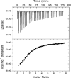
ITC curve of binding of haloxyfop-R to M. tuberculosis AccD6.
FIG 9
FIG 9
(A) Superimposition of M. tuberculosis AccD6 and AccD5. Cα RMSD, 1.6 Å. AccD6 is in blue and AccD5 (PDB code 2A7S) is in green. (B) Comparison of stacking interactions between M. tuberculosis AccD6 (green) and the yeast CT domain (red). (C) Comparison of the haloxyfop trifluoro methyl binding site. M. tuberculosis AccD6 site 1 is in green, and the yeast CT domain is in red. (D) Comparison of the hydrogen bonding at the acid position of haloxyfop. M. tuberculosis AccD6 site 1 is in green, and the yeast CT domain is in red. Yeast residue numbering is in italics.

References

    1. Wakil SJ, Stoops JK, Joshi VC. 1983. Fatty acid synthesis and its regulation. Annu. Rev. Biochem. 52:537–579. 10.1146/annurev.bi.52.070183.002541 - DOI - PubMed
    1. Cronan JE, Jr, Waldrop GL. 2002. Multi-subunit acetyl-CoA carboxylases. Prog. Lipid Res. 41:407–435. 10.1016/S0163-7827(02)00007-3 - DOI - PubMed
    1. Tanabe T, Wada K, Okazaki T, Numa S. 1975. Acetyl-coenzyme-A carboxylase from rat liver. Subunit structure and proteolytic modification. Eur. J. Biochem. 57:15–24 - PubMed
    1. Cole ST, Brosch R, Parkhill J, Garnier T, Churcher C, Harris D, Gordon SV, Eiglmeier K, Gas S, Barry CE, Tekaia F, Badcock K, Basham D, Brown D, Chillingworth T, Conner R, Davies R, Devlin K, Feltwell T, Gentles S, Hamlin N, Holroyd S, Hornsby T, Jagels K, Krogh A, McLean J, Moule S, Murphy L, Oliver K, Osborne J, Quail MA, Rajandream MA, Rogers J, Rutter S, Seeger K, Skelton J, Squares R, Squares S, Sulston JE, Taylor K, Whitehead S, Barrell BG. 1998. Deciphering the biology of Mycobacterium tuberculosis from the complete genome sequence. Nature 393:537–544. 10.1038/31159 - DOI - PubMed
    1. Lin TW, Melgar MM, Kurth D, Swamidass SJ, Purdon J, Tseng T, Gago G, Baldi P, Gramajo H, Tsai SC. 2006. Structure-based inhibitor design of AccD5, an essential acyl-CoA carboxylase carboxyltransferase domain of Mycobacterium tuberculosis. Proc. Natl. Acad. Sci. U. S. A. 103:3072–3077. 10.1073/pnas.0510580103 - DOI - PMC - PubMed

Publication types

MeSH terms