. 2013 Apr;69(Pt 4):603-16.
doi: 10.1107/S0907444913000024. Epub 2013 Mar 14.

Structural insights into lipid-dependent reversible dimerization of human GLTP

Affiliations

Structural insights into lipid-dependent reversible dimerization of human GLTP

Valeria R Samygina et al. Acta Crystallogr D Biol Crystallogr. 2013 Apr.

Abstract

Human glycolipid transfer protein (hsGLTP) forms the prototypical GLTP fold and is characterized by a broad transfer selectivity for glycosphingolipids (GSLs). The GLTP mutation D48V near the `portal entrance' of the glycolipid binding site has recently been shown to enhance selectivity for sulfatides (SFs) containing a long acyl chain. Here, nine novel crystal structures of hsGLTP and the SF-selective mutant complexed with short-acyl-chain monoSF and diSF in different crystal forms are reported in order to elucidate the potential functional roles of lipid-mediated homodimerization. In all crystal forms, the hsGLTP-SF complexes displayed homodimeric structures supported by similarly organized intermolecular interactions. The dimerization interface always involved the lipid sphingosine chain, the protein C-terminus (C-end) and α-helices 6 and 2, but the D48V mutant displayed a `locked' dimer conformation compared with the hinge-like flexibility of wild-type dimers. Differences in contact angles, areas and residues at the dimer interfaces in the `flexible' and `locked' dimers revealed a potentially important role of the dimeric structure in the C-end conformation of hsGLTP and in the precise positioning of the key residue of the glycolipid recognition centre, His140. ΔY207 and ΔC-end deletion mutants, in which the C-end is shifted or truncated, showed an almost complete loss of transfer activity. The new structural insights suggest that ligand-dependent reversible dimerization plays a role in the function of human GLTP.

Keywords: GLTP fold; glycolipid transfer protein; lipid-mediated homodimerization; selectivity; sulfatides.


Figures
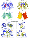
Figure 1
Figure 1
Dimeric arrangement, flexibility and intermolecular contacts in wild-type GLTP (wtGLTP) and the D48V mutant complexed with the short-acyl-chain sulfatide 12:0 monoSF. (a, b) Open dimer conformation in wtGLTP (a) versus the locked dimer of D48V (b). Protein chains are coloured blue for wtGLTP and gold for the D48V mutant. Sulfatide atoms are coloured red, blue and green for oxygen, nitrogen and sulfur, respectively, while C atoms are coloured pink for the lipid bound to wtGLTP and blue for the lipid bound to the D48V mutant. The eight α-helices of the GLTP fold are labelled α1–α8. The values 80° and 65° indicate the mutual inclination of two α2 helices in the dimeric structure of wtGLTP versus D48V-GLTP. (c) Comparative superimposition of ‘open’ and ‘closed’ dimer conformations for different wtGLTP crystal forms (referred to as WT3 and WT2). Protein Cα chains are coloured blue and green for different dimers. The monomers on the left are superimposed to highlight the range of openness of the monomer on the right for the different dimers. Blue and green semi-transparent solid shapes additionally highlight the superimposition of the left monomers and the range of openness of the right monomer for the different dimers. Ligand molecules are omitted for clarity. (d) Schematic highlighting the closed and locked conformation of the D48V dimers. Complexes with 12:0 monoSF and with 12:0-diSF are shown by solid shapes coloured yellow and red–brown, respectively, with α2, α6 and α8 indicating selected helices and the rectangle denoting the hydrophobic core that ‘locks’ the dimer. For the superposition of Cα chains, see Supplementary Fig. S1(c). (eg) Protein–protein′ dimeric contact regions shown by comparative superimposition of wtGLTP and D48V: overall view (e) and two zones of expanded contacts in the locked dimer (f, g). Superpositioning details and colour codes are as in (a) and (c). Wide dashed bands show expanded contacts. Solid straight lines are conditional axes of α2 helices, while dashed lines with values indicate the midpoint distances between them (t distances in Table 3 ▶).
Figure 2
Figure 2
Various conformations of 12:0-monoSF in wtGLTP and GLTP mutants. (a, b) Comparative superpositions of 12:0 monoSF with 24:1 monoSF as bound to wtGLTP (a) versus D48V (b). Protein moieties are omitted after being used for superimpositioning. Sulfatide atoms are coloured red, blue and green for oxygen, nitrogen and sulfur, respectively. 12:0-monoSF C atoms and extraneous hydrocarbons found in the hydrophobic pocket are coloured magenta in wtGLTP and cyan in D48V; 24:1 monoSF C atoms are coloured light magenta and light cyan, respectively. (c) Comparative superposition of 12:0 monoSF conformations in wtGLTP and D48V. Colour codes are as in (a) and (b). (df) Dimeric arrangements of the sphingosine-out modes in wtGLTP (d), D48V (e) and A47D/D48V (f). Colour codes are as in (a) and (b), except for the C atoms of A47D/D48V, which are shown in white. Red and green semitransparent rectangles highlight the sphingosine chains of the left and right monomers, respectively.
Figure 3
Figure 3
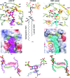
Disulfatide binding to wtGLTP and the D48V mutant. (a) 3,6-O-Disulfo-Gal headgroup in the wtGLTP recognition centre. Dashed lines indicate hydrogen bonds. Disulfatide atoms are coloured red, blue, green and magenta for oxygen, nitrogen, sulfur and carbon, respectively. Protein Cα backbone and side-chain C atoms are coloured silver and gold, respectively. The grey circle labelled W1 or W indicates the conserved water molecule. S1 and S2 (pink rectangles) are 3-O- and 6-O-sulfo groups, respectively. (b) Chemical structure of N-lauroyl-3,6-O-disulfo-galactosylceramide. (c) 3,6-O-Disulfo-Gal headgroup in the recognition centre of the D48V mutant. Colour codes and designations are as in (a), except for ligand C atoms, which are coloured cyan. The mutated residue 48 is shown in a red circle; the black arrow points out the different conformation of S2 compared with that in wtGLTP (a). (de) Electrostatic surface view (blue, positive; red, negative; grey, neutral) of the GLTP recognition centre in the wild-type protein (d) and the D48V mutant (e) occupied by a disulfatide molecule shown in stick representation within a space-filled semitransparent shape with green-coloured sulfo groups. Colour codes are as in (a) and (b). The mutated residue is shown in a red circle; arrows point out the ‘empty’ space (filled by water molecules) resulting from the D48V mutation and the conformational change of the S2 group promoting the movement of the C-end. (f, h) Dimeric arrangements of 12:0-diSF in wtGLTP (f) and D48V (h), with the ligand in ball-and-stick representation. Colour codes are as in Figs. 2 ▶(d) and (e). (g) Superimposed disulfatide molecules as bound to D48V (cyan) versus wtGLTP (magenta).
Figure 4
Figure 4
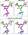
Conformational adaptability of disulfatide headgroups. (a) Superimposed ligand conformations in open and closed dimers (crystal forms 1 and 2, respectively) of wtGLTP. Protein moieties were superposed and skipped. Disulfatide molecules are shown in stick representation; colour codes for O, N and S atoms are red, blue and green, respectively. C atoms are coloured magenta for crystal form 1 and pink for crystal form 2. (b) Disulfatide conformations in two crystal forms (1 and 4; Tables 1 ▶ and 2 ▶) of D48V. C atoms are coloured cyan for crystal form 1 and light cyan for crystal form 4. Colour codes for other atoms are as in (a). (c, d) Disulfatide in the open dimer of wtGLTP versus the two conformations of disulfatide shown in (b).
Figure 5
Figure 5
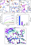
Involvement of the protein C-end in the network of interactions supporting residue His140 in human GLTP. (a) The His140 side chain makes a key hydrogen bond to the ceramide amide group and multiple contacts with the environment. Ligand and amino acids are shown in stick representation. C atoms are coloured magenta in the lipid, silver in the amino-acid residues surrounding His140 and orange in the partner monomer. Colour codes for O and N atoms are red and blue, respectively. Dashed lines indicate hydrogen bonds, while zigzags are close van der Waals contacts. The C-terminal residue Val209 simultaneously contacts His140 and Pro40* from the partner. (b) The main chain of residue His140 supported by the network of hydrogen bonds involving the C-end of the protein main chain. Designations are as in (a). The circled part of the network highlights the C-end contribution. (c) Disposition of the circled part of the network in proximity to the recognition centre of hsGLTP (the Val209 side chain is skipped). Colour codes are as in (a). (d, e) Transfer activity assays for wtGLTP and mutants. (d) Transfer of the AV-glycolipid by wt-GLTP (blue), Y207L (magenta), Δ207 (green) or D48V/ΔC-end (orange) as a function of time. The increase in fluorescence emission at 415 nm (AV emission) occurs because of decreased Förster resonance energy transfer when AV-glycolipid is removed from donor vesicles containing 3-perylenoyl PC and is transported to POPC acceptor vesicles (see §2 for details). The AV signal change does not occur in the absence of POPC acceptor vesicles. (e) Transfer activity of GLTP by D48V, Y207L mutations or Y207/C-end deletions. The initial rates of AV-glycolipid departure from the donor vesicles are shown. (f) Expanded view of interactions shown in (b) for the open dimeric arrangement of wtGLTP complexed with monoSF (magenta) versus the locked dimer of the D48V mutant complexed with diSF (cyan). Parts of the partner protein monomer contacting the C-end are highlighted by additional sphere representations in an appropriate colour.

References

    1. Abe, A. (1990). J. Biol. Chem. 265, 9634–9637. - PubMed
    1. Airenne, T. T., Kidron, H., Nymalm, Y., Nylund, M., West, G., Mattjus, P. & Salminen, T. A. (2006). J. Mol. Biol. 355, 224–236. - PubMed
    1. Bahadur, R. P. & Zacharias, M. (2008). Cell. Mol. Life Sci. 65, 1059–1072. - PMC - PubMed
    1. Brown, R. E. (1992). Biochim. Biophys. Acta, 1113, 375–389. - PMC - PubMed
    1. Brown, R. E. & Mattjus, P. (2007). Biochim. Biophys. Acta, 1771, 746–760. - PMC - PubMed

Publication types