. 2013 Jan 17;152(1-2):183-95.
doi: 10.1016/j.cell.2012.12.025.

The myosin chaperone UNC-45 is organized in tandem modules to support myofilament formation in C. elegans

Affiliations

The myosin chaperone UNC-45 is organized in tandem modules to support myofilament formation in C. elegans

Linn Gazda et al. Cell. .

Abstract

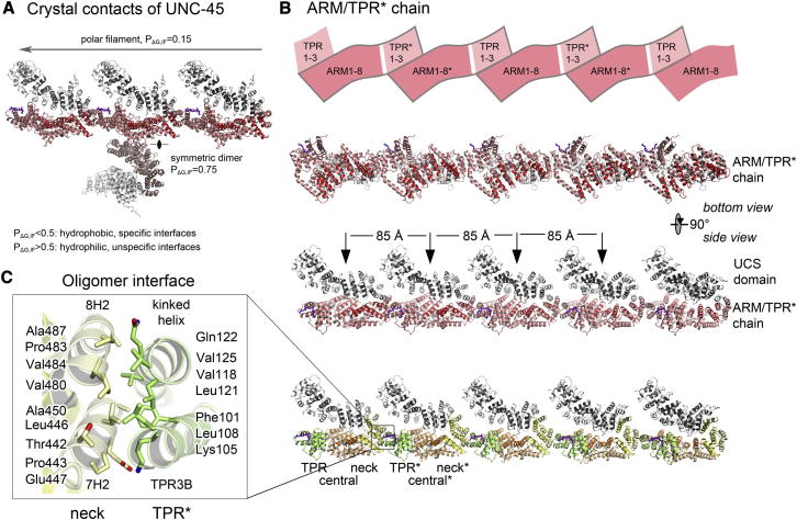
The UCS (UNC-45/CRO1/She4) chaperones play an evolutionarily conserved role in promoting myosin-dependent processes, including cytokinesis, endocytosis, RNA transport, and muscle development. To investigate the protein machinery orchestrating myosin folding and assembly, we performed a comprehensive analysis of Caenorhabditis elegans UNC-45. Our structural and biochemical data demonstrate that UNC-45 forms linear protein chains that offer multiple binding sites for cooperating chaperones and client proteins. Accordingly, Hsp70 and Hsp90, which bind to the TPR domain of UNC-45, could act in concert and with defined periodicity on captured myosin molecules. In vivo analyses reveal the elongated canyon of the UCS domain as a myosin-binding site and show that multimeric UNC-45 chains support organization of sarcomeric repeats. In fact, expression of transgenes blocking UNC-45 chain formation induces dominant-negative defects in the sarcomere structure and function of wild-type worms. Together, these findings uncover a filament assembly factor that directly couples myosin folding with myofilament formation.


Figures

None
Graphical abstract
Figure 1
Figure 1
Structure of UNC-45 (A) Ribbon presentation of the UNC-45 protomer showing its domain architecture. The numbering of the TPR and ARM repeats is given with helix H3 of each ARM repeat being highlighted. The cocrystallized Hsp90 peptide is shown in stick mode (lilac). Dashed lines represent flexible loops comprising residues 508–524 and 608–617. (B) Orthogonal view of the UNC-45 protomer highlighting the two distinct faces of the UCS domain. (C) Sequence alignment of the UNC-45 proteins from C. elegans (Ce; Q09981), Drosophila melanogaster (Dm; Q9VHW4), Homo sapiens (Hs; E1P642), and Danio rerio (Dr; Q6DGE9). The nomenclature of the TPR and ARM helices is given. With the exception of the UCS signature motif (loop L602–630) colored in magenta, the colors of the secondary structure elements reflect the domain colors. Dotted lines represent flexible loops that are not defined by electron density. Positions of UNC-45 temperature-sensitive (ts) mutations are indicated by asterisks. Functionally important residues are marked with a triangle (dark gray, oligomer interface inner core; light gray, oligomer interface outer residues; green, binding to Hsp90 C terminus; blue, TPR central interface). Red residues are strictly conserved in all four sequences; boxed residues are highly conserved. The overall sequence identity between the CeUNC-45 and the other listed UNC-45 proteins is about 30% (48% similarity). See also Figure S1 and Table S1.
Figure 2
Figure 2
Interaction of UNC-45 with Its Partner Chaperones Hsp70 and Hsp90 (A) Representative ITC data recorded upon mixing wild-type and K82E UNC-45 with the Hsp70/90 C-terminal peptides. Raw data (top) and binding isotherm derived from the integrated heat (bottom) are shown. Calculated KD values are given. (B) SEC/SDS-PAGE analysis reveals complex formation of UNC-45 with Hsp90 (left) and Hsp70 (right). (C) Binding mode of the Hsp90 C terminus (lilac) to the TPR domain (green). Interacting residues of the TPR domain are labeled. (D) Overlay of the Hsp70 (blue) and Hsp90 (lilac) peptide structures bound to the TPR domain (green). Close-up view of the Hsp90 peptide (top), Hsp70 peptide (bottom), and interacting TPR domain residues overlaid with the 2Fo – Fc omit electron density (Hsp90: 2.9 Å resolution, contoured at 1.1σ; Hsp70: 3.6 Å, contoured at 1.1σ). Interactions with Hsp90 that are not formed with Hsp70 are highlighted with an asterisk. See also Figure S2.
Figure 3
Figure 3
UNC-45 Interaction with Myosin (A) Ribbon model of the UCS domain highlighting mutations (magenta) used to address the myosin binding of UNC-45. To illustrate the position of substrate, a peptide ligand (green) of a superimposed β-catenin molecule was transferred to the UNC-45 fold. The zoom-up windows provide a detailed view on the Y750W and N758Y mutations, the functionally related residues in β-catenin (PDB code 1i7w, with bound E-cadherin peptide shown as stick model) and the Δ602-630 deletion expected to interrupt the UCS canyon. (B) To estimate the rescue of the uncoordinated (unc) phenotype, young adult unc-45(m94) worms expressing the indicated UNC-45 variants were grown at 25°C and body bends counted (two independent transgenic lines per mutation, SE of the mean indicated). (C) Immunostaining of unc-45(m94) mutant worms grown at 25°C expressing Y750W, N758Y, or Δ602–630 UNC-45. (D) Protein-protein interactions undergone by wild-type and mutant UNC-45 (FLAG tag indicated by asterisk). The UNC-54 myosin and Hsp90 were coimmunoprecipitated from cell lysates of the indicated unc-45(m94) mutant worms expressing different UNC-45 variants grown at 25°C. Protein interactions were evaluated by western blot analysis using UNC-54-, Hsp90-, and UNC-45-specific antibodies. See also Figure S3 and Table S2.
Figure 4
Figure 4
Structural Organization of the UNC-45 Multimer (A) Crystal contacts observed in the unit cell of C. elegans UNC-45. Respective PΔG,IF values reflect the likelihood that the resultant dimer or filament represents a crystallographic artifact. (B) Architecture of the UNC-45 oligomer. The ARM/TPR building block is schematically shown as enclosed box in the top panel (molecular neighbors distinguished by asterisks). The ribbon presentations in the bottom panels provide an orthogonal view on the UNC-45 chain and illustrate that TPR, central, and neck domain form the backbone of the assembly, whereas the UCS domain projects away offering myosin-binding sites in a regular array (indicated by arrows). (C) Close-up view of the oligomer interface showing the composite four-helix bundle. Involved helices and residues are labeled. See also Figure S4 and Table S3.
Figure 5
Figure 5
In Vitro Validation of UNC-45 Chains (A) SDS-PAGE analysis of the photocrosslinking reaction upon pBpa452 (Q452B, indicated by “X”) activation. Samples of wild-typex, L121Wx, V480R/V484Rx, and ΔTPRx UNC-45 were exposed for 15 and 30 min to UV illumination. Schematic representations of the introduced mutations and resultant crosslinks are indicated. Specifically linked oligomers (as deduced by MS) are marked by arrows, whereas dimers resulting from unspecific crosslinking are labeled with a circle. (B) SEC elution profile and corresponding SDS-PAGE of the wild-typex sample illustrating formation of 2–5 meric UNC-45 chains. (C) MS2 spectrum of the crosslinked peptide identified in the marked Q452B oligomers. The inset shows the observed product ions mapped onto the sequence of the crosslinked peptide, the isotopic distribution of the crosslinked peptide, its mass-to-charge ratio (m/z), its charge (3+), and its monoisotopic mass value (m). Δmass, difference between the expected and measured masses; R, resolution of the measurement; B, pBpa; M, oxidized methionine. (D) In the crystal structure of the UNC-45 chain, the identified crosslinked residues Lys131-Ile132 are juxtaposed to the pBpa452 of the molecular neighbor. Because these residues were exclusively identified in the UNC-45 2 mer, 3 mer, 4 mer, and 5 mer, the MS data are consistent with the illustrated protein chains. The close-up window illustrates the molecular details of the TPR-ARM oligomer interface highlighting the mutated (black) and crosslinked (magenta) residues. See also Figure S5.
Figure 6
Figure 6
Functional Analysis of UNC-45 Oligomerization Mutants In Vivo (A) Ribbon plot providing the close-up view of the oligomer interface with the mutated residues Val480, Val484, and Leu121 shown as orange spheres and the ΔTPR enclosed by a box. (B–E) To estimate the rescue of the uncoordinated (unc) phenotype, young adult unc-45(m94) worms expressing the indicated UNC-45 variants were grown at 25°C. Immunostaining of UNC-45 and UNC-54 (myosin), body bend counts, the ability of UNC-45-FLAG variants to coIP UNC-54 (myosin), and sarcomeric A band quantification are shown, respectively. (F–H) The dominant-negative effect of expressing different UNC-45-FLAG versions in unc-119/unc-45(wt) worms was assayed by body bend counts, A band staining, and quantification, respectively. The number of A bands (labeled with anti-MHC A) is given per body wall muscle cell (indicated by dotted line), with all analyzed cells located in the same area between pharynx and vulva. All values are mean ± SEM. See also Figure S6, Table S2, and Table S4.
Figure 7
Figure 7
Model of UNC-45-Promoted Myosin Assembly (A) UNC-45 composes a myosin folding complex. (Top) UNC-45 (different domains indicated), its oligomers, Hsp70 (blue), and Hsp90 (lilac) represent the basic building blocks for the myosin multichaperone complex. (Bottom) UNC-45 chains compose a molecular scaffold that allows the simultaneous binding of Hsp70, Hsp90, and myosin, thus mediating the interactions between unfolded myosin and its cognate chaperones. Different lengths of the Hsp70/90 C-terminal linkers and resultant activity radii when bound to UNC-45 are indicated. (B) UNC-45 enforces the folding of myosin in regular spacing. In the relaxed state of the muscle, dimeric myosin heads project with a periodicity of 145 Å from the coiled-coil backbone of the thick filament. The observed periodicity between UNC-45 tandem modules (170 Å) fits well to the observed spacing between protomers of adjacent myosin pairs (110 to 220 Å, derived from PDB code 3dtp), as schematically shown in the enlarged window (myosin in green with orange circles symbolizing equal segments in the asymmetric dimer). Owing to its patterning function, UNC-45 could directly link myosin assembly with myosin folding.
Figure S1
Figure S1
Domain Organization and Interfaces of CeUNC-45, Related to Figure 1 (A) Ribbon model and folding topology of UNC-45 illustrating its domain architecture and the used helix nomenclature (TPR: green; central: orange; neck: yellow; UCS: gray). Each TPR motif is made of two α helices A and B, whereas the ARM repeats are composed of three helices H1, H2 and H3. The helices H2 and H3 (shown as circles) of neighboring repeats are arranged in parallel constituting the core of the individual domains, whereas the relatively short H1 helices (shown as bars, not labeled), which are perpendicularly arranged to H2 and H3, orient adjacent ARM repeats to each other. Disordered loops are indicated by a dotted line. (B) The central domain serves as a scaffolding unit that arranges TPR and UCS domain. The two close-up views illustrate the corresponding UNC-45 domain interfaces (polar interactions represented as dotted lines). Upper panel: Interface of central and UCS domain. Lower panel: Interface of central and TPR domain highlighting several short-distanced salt-bridges formed between Asp62-Lys269, Arg53-Asp279, Lys72-Glu281 and Glu29-Lys277. (C) Sequence alignment of the UCS myosin-binding domain of CeUNC-45, Saccharomyces cerevisiae (Sc) She4, Schizosaccharomyces pombe (Sp) Rng3, Podospora anserina (Pa) CRO1, the founding members of the UCS protein family. Helices identified in the CeUNC-45 structure are indicated. Conserved residues are highlighted (red, strictly conserved; boxed, highly conserved) and reveal the extremely high conservation of the functionally important 13H3 helix pointing to a common mechanism in myosin binding. The signature motif in the UCS domain is colored in magenta. The sequence identity (similarity) of the UCS domain between CeUNC-45 and She4, Rng3 and CRO1 is 15% (26%), 21% (34%) and 23% (36%), respectively.
Figure S2
Figure S2
Details of Hsp70 and Hsp90 Binding to UNC-45, Related to Figure 2 (A) Analytical SEC and SDS-PAGE of UNC-45, Hsp70 and Hsp90. Left panel: UNC-45 and equal concentrations of Hsp70 and Hsp90 full-length proteins. Right panel: UNC-45 and Hsp90 in the presence of a decapeptide representing the C terminus of Hsp70. (B) Analytical SEC and SDS-PAGE of UNC-45 and its partner chaperones Hsp70 or Hsp90 lacking their C-terminal motives IEEVD and MEEVD, respectively. (C) Molecular surface of UNC-45 with mapped conservation pattern (red, highly conserved; yellow, less conserved) and bound Hsp90 peptide (lilac). The close-up view of the TPR domain shows the strictly conserved binding site for the Hsp90 peptide. (D) UNC-45 was co-crystallized with a decapeptide representing either the Hsp70 or the Hsp90 C terminus. Left panel: Stereo view of the TPR domain (green) and the co-crystallized Hsp70 (blue) and Hsp90 (lilac) peptides shown in stick mode. The final model is overlaid with the respective 2Fo-Fc omit electron density calculated without ligand (complex with Hsp90: 2.9 Å resolution, contoured at 1.1σ; Hsp70: 3.6 Å resolution, contoured at 1.1σ). Interacting residues are labeled. To describe the specific contacts with UNC-45, we will focus on the interaction with the Hsp90 peptide, which is numbered in descending order with the C-terminal Asp0 preceded by Val-1, Glu-2, Glu-3, Met-4, Arg-5, Ser-6 and Ala-7. The Hsp90 C terminus is tethered in a bent conformation to the TPR domain undergoing multiple main-chain and side-chain interactions with highly conserved UNC-45 residues. Contacting residues of the TPR domain are highlighted in green and are labeled. Contacts to the backbone are mediated by Lys82 and Arg86 that bind to the carbonyl oxygen of Glu-2, by Asp114 that interacts with the amine nitrogen of Met-4 and finally by Arg12, Asn16 and Asn52 that compose a positively charged pocket to accommodate the carboxy-terminus of Hsp90. Notably, the geometry of the three backbone anchor points favors binding of the Hsp90 peptide in bent conformation. The observed turn structure is further stabilized by an internal hydrogen bond between the side chains of Glu-2 and Ser-6. All remaining side-chains of the Hsp90 peptide are engaged in interactions with highly conserved TPR residues ensuring the tight and specific binding of the Hsp90 C-terminal peptide. The carboxylate group of Asp0 is tightly bound by Arg51 and Lys82, the Val-1 side chain is accommodated in a hydrophobic pocket lined by residues Val19, Asn52 and Met55, Glu-3 forms a short-distanced salt-bridge with the amino group of Lys59 and the side-chain of Met-4 protrudes into a deep hydrophobic pocket bordered by Val81, Lys82, Phe85 and Ile117. Right panel: Eight residues of the Hsp90 peptide ligand (ASRMEEVD lilac) and ten residues of the Hsp70 peptide ligand (AGGPTIEEVD blue) are well-defined by electron density revealing their precise binding mode. Additional flexible residues at the C-termini of Hsp70 (∼20 amino acids) and Hsp90 (∼5 amino acids) are shown schematically as dotted lines.
Figure S3
Figure S3
Potential Myosin-Binding Site of the UCS Domain, Related to Figure 3 (A) Closest structural homologs, as revealed by a DALI search, are listed for the UCS domain. (B) Left panel: Structural alignment of the UCS domain of UNC-45 (gray) with the ARM domain of β-catenin (violet, PDB code: 1i7x). Right panel: To identify the myosin binding site of UNC-45, peptide ligands (green) were extracted from aligned β-catenin crystal structures and mapped on the UCS domain. H3 helices of the UCS domain are highlighted with helix 13H3 and the UCS loop shown in magenta. (C) Left panel: Molecular surface of the UCS domain with mapped electrostatic potential and aligned β-catenin peptide ligand. Right panel: Molecular surface of the UCS domain with mapped conservation pattern and aligned β-catenin peptide ligand. The illustration highlights the hydrophobic surface and the conservation of the UCS canyon.
Figure S4
Figure S4
Crystal Packing of CeUNC-45, DmUNC-45, and She4, Related to Figure 4 (A) Structural alignments of CeUNC-45 (gray) with DmUNC-45 (PDB code: 3now, magenta, top) and She4 (PDB code: 3opb, blue, bottom panel). The 602-630 loop of CeUNC-45 extends as unfolded segment into the protein periphery, whereas in the crystal structure of DmUNC-45, this loop folds back on the UCS domain filling partly the extended central canyon (shown in orange). We presume that the different domain organization and oligomeric states observed in the crystal structures of DmUNC-45 and She4 are due to different crystal contacts and the introduction of multiple mutations for protein crystallization, respectively. (B) CeUNC-45 was crystallized in space group P6122 having a single molecule in the asymmetric unit (unit cell dimensions are indicated). Six UNC-45 dimers related by a crystallographic 2-fold axis are sitting in a 61-screw axis on top of each other thus yielding the long c-axis of 815 Å. Each of the 12 molecules undergoes longitudinal interactions with their crystallographic neighbors (shown for top molecule) resulting in polar UNC-45 chains that extend with a 85 Å periodicity throughout the crystal, perpendicularly to the long crystallographic c-axis. In contrast to the filament interface, the crystallographic dimer is largely stabilized by polar interactions as shown in the close-up window. (C) DmUNC-45 was crystallized in space group P23 with a single subunit in the asymmetric unit. Owing to crystal symmetry, twelve molecules associate via their UCS domains forming a hollow sphere that is surrounded by six couples of central domains. Side-by-side packing of the DmUNC-45 spheres generates a wide-meshed crystal lattice with huge holes, into which the flexible TPR domains (indicated by green spheres) protrude. Whether the en-bloc flexibility of the TPR domain is functionally relevant or influenced by crystal packing remains to be shown. (D) She4, a yeast UCS protein that is proposed to promote actin-myosin interactions (Shi and Blobel, 2010), was crystallized upon introducing 13 point mutations and deleting loop 343-358 (indicated by red circles). In the crystal lattice, two She4 molecules associate to generate an S-shaped dimer that is stabilized by the N-terminal helix binding to the neck domain (ARM repeats 8-9) of a molecular neighbor. It still needs to be experimentally shown that the crystallographic dimer is functionally relevant.
Figure S5
Figure S5
Concentration Dependency of UNC-45 Oligomer Formation, Related to Figure 5 (A) SDS-PAGE analysis of the photo-crosslinking reaction upon pBpa452 activation. Samples of wild-type UNC-45 Q452 pBpa exposed to UV illumination for 15 and 30 min at three different concentrations are shown. The unspecific dimer is marked with a circle. (B) Quantification of the crosslinked fraction at 30 min. Percentage of dimer (lilac), trimer (red) and tetramer (green) are shown for the different concentrations. 100% crosslink at 100 μM concentration is used as reference.
Figure S6
Figure S6
Analysis of UNC-45 Oligomer Formation Mutants, Related to Figure 6 (A– C) (A) Immunostaining of MHC A in unc-45(m94) worms expressing either wild-type UNC-45-FLAG or the indicated UNC-45-FLAG oligomer formation mutants (FLAG-tag indicated by asterisk). Single cells are outlined by a dotted line. Total UNC-54 levels in unc-45(m94) worms (B) and unc-119/unc-45(wt) worms (C) expressing different UNC-45-FLAG versions were determined by anti-UNC-54 Western Blot analysis. An anti-tubulin Western Blot is shown as loading control. (D) UNC-54 myosin was coimmunoprecipitated with the indicated UNC-45-FLAG versions from unc-119/unc-45(wt) worms expressing the indicated UNC-45-FLAG variants. Proteins were detected by Western Blot analysis. (E) Molecular surface representation of UNC-45 (gray) and an aligned β-catenin peptide ligand (green) in stick mode. The site-specific oligomer formation mutants L121W and V480R/V484R are highlighted in orange.

References

    1. Ao W., Pilgrim D. Caenorhabditis elegans UNC-45 is a component of muscle thick filaments and colocalizes with myosin heavy chain B, but not myosin heavy chain A. J. Cell Biol. 2000;148:375–384. - PMC - PubMed
    1. Barral J.M., Bauer C.C., Ortiz I., Epstein H.F. Unc-45 mutations in Caenorhabditis elegans implicate a CRO1/She4p-like domain in myosin assembly. J. Cell Biol. 1998;143:1215–1225. - PMC - PubMed
    1. Barral J.M., Hutagalung A.H., Brinker A., Hartl F.U., Epstein H.F. Role of the myosin assembly protein UNC-45 as a molecular chaperone for myosin. Science. 2002;295:669–671. - PubMed
    1. Bernick E.P., Zhang P.J., Du S. Knockdown and overexpression of Unc-45b result in defective myofibril organization in skeletal muscles of zebrafish embryos. BMC Cell Biol. 2010;11:70. - PMC - PubMed
    1. Craig R., Woodhead J.L. Structure and function of myosin filaments. Curr. Opin. Struct. Biol. 2006;16:204–212. - PubMed

Supplemental References

    1. Abrahams, J.P., and Leslie, A.G. (1996). Methods used in the structure determination of bovine mitochondrial F1 ATPase. Acta Crystallogr. D Biol. Crystallogr. 52, 30–42. - PubMed
    1. Abramoff, M.D., Magalhães, P.J., and Ram, S.J. (2004). Image processing with ImageJ. Biophotonics Intl. 11, 36–42.
    1. Adams, P.D., Grosse-Kunstleve, R.W., Hung, L.W., Ioerger, T.R., McCoy, A.J., Moriarty, N.W., Read, R.J., Sacchettini, J.C., Sauter, N.K., and Terwilliger, T.C. (2002). PHENIX: building new software for automated crystallographic structure determination. Acta Crystallogr. D Biol. Crystallogr. 58, 1948–1954. - PubMed
    1. Ben-Zvi, A., Miller, E.A., and Morimoto, R.I. (2009). Collapse of proteostasis represents an early molecular event in Caenorhabditis elegans aging. Proc. Natl. Acad. Sci. USA 106, 14914–14919. - PMC - PubMed
    1. Brenner, S. (1974). The genetics of Caenorhabditis elegans. Genetics 77, 71–94. - PMC - PubMed

Publication types

MeSH terms

Substances

LinkOut - more resources