. 2013 Mar;5(3):212-20.
doi: 10.1038/nchem.1565. Epub 2013 Feb 3.

Volume-conserving trans-cis isomerization pathways in photoactive yellow protein visualized by picosecond X-ray crystallography

Affiliations

Volume-conserving trans-cis isomerization pathways in photoactive yellow protein visualized by picosecond X-ray crystallography

Yang Ouk Jung et al. Nat Chem. 2013 Mar.

Abstract

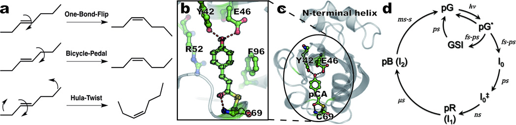
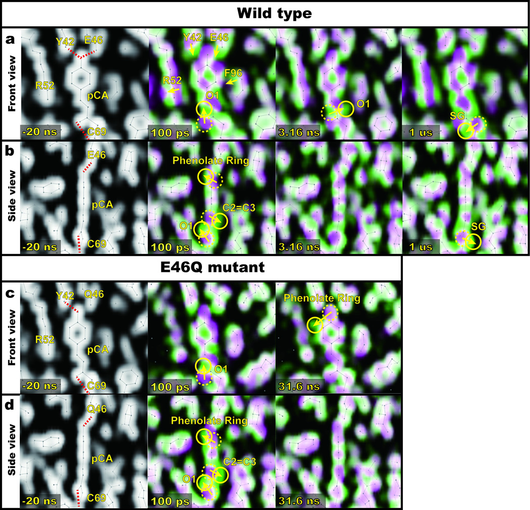
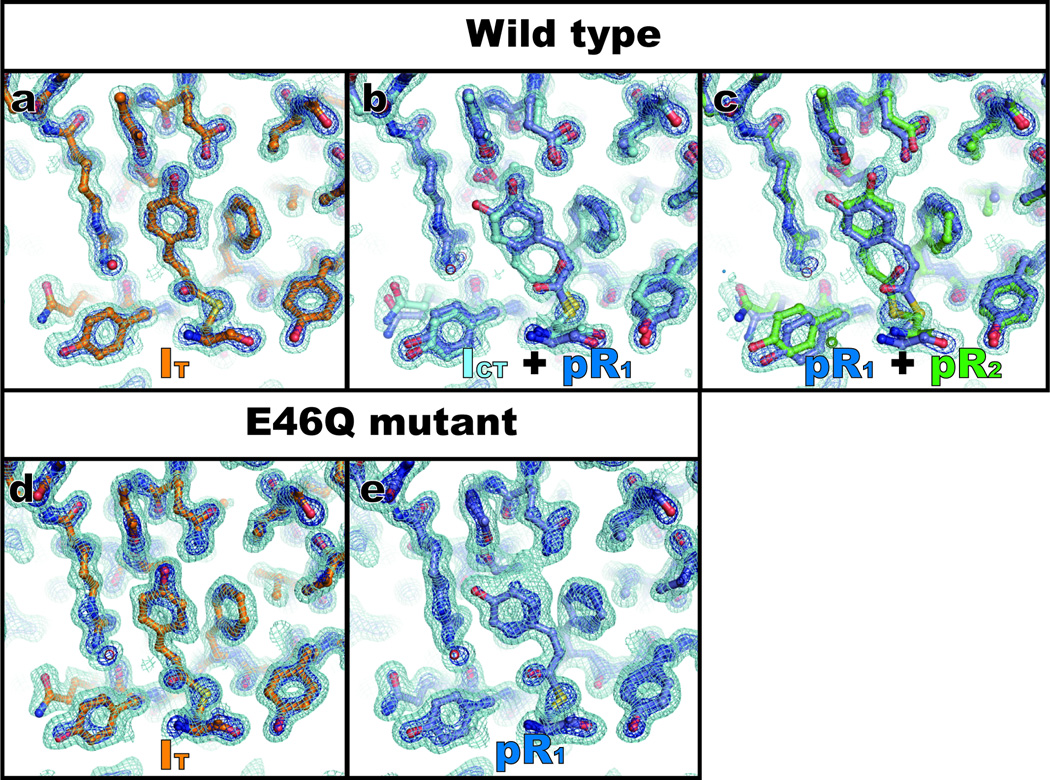
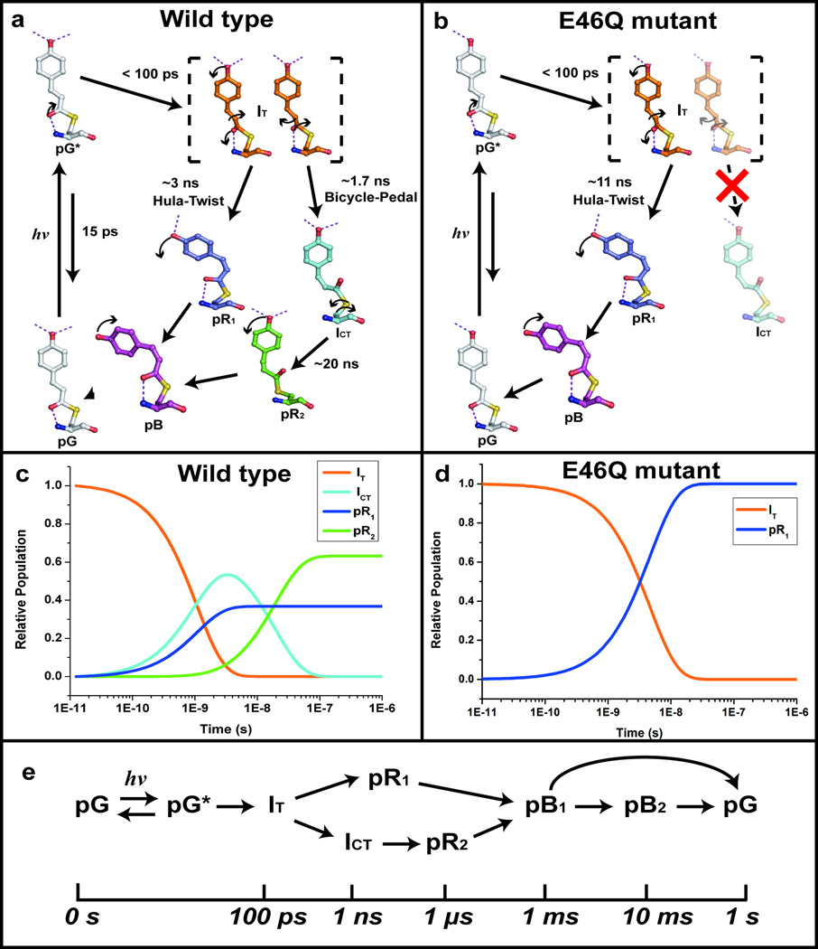
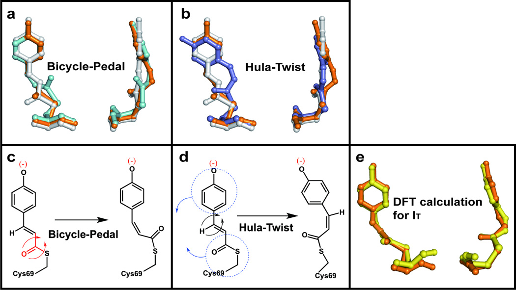
Trans-to-cis isomerization, the key reaction in photoactive proteins, usually cannot occur through the standard one-bond-flip mechanism. Owing to spatial constraints imposed by a protein environment, isomerization probably proceeds through a volume-conserving mechanism in which highly choreographed atomic motions are expected, the details of which have not yet been observed directly. Here we employ time-resolved X-ray crystallography to visualize structurally the isomerization of the p-coumaric acid chromophore in photoactive yellow protein with a time resolution of 100 ps and a spatial resolution of 1.6 Å. The structure of the earliest intermediate (I(T)) resembles a highly strained transition state in which the torsion angle is located halfway between the trans- and cis-isomers. The reaction trajectory of I(T) bifurcates into two structurally distinct cis intermediates via hula-twist and bicycle-pedal pathways. The bifurcating reaction pathways can be controlled by weakening the hydrogen bond between the chromophore and an adjacent residue through E46Q mutation, which switches off the bicycle-pedal pathway.


Figures

Figure 1
Figure 1
Isomerization mechanisms and overview of photoactive yellow protein. (a) Schematic description of three isomerization mechanisms discussed in this work. (b) Close-up of the pCA chromophore and neighboring residues; dashed lines denote hydrogen-bond interactions. Carbon, oxygen, sulfur, and nitrogen atoms are shown in green, red, yellow, and blue, respectively. (c) Structure of the protein (ribbon) and the pCA (ball-and-stick) in the chromophore binding pocket. (d) Photocycle and corresponding kinetics, as derived from time-resolved spectroscopy measurements at ambient temperature–,,.
Figure 2
Figure 2
Time-resolved electron density maps of pCA in the chromophore binding pocket. For clarity, only 100 ps, 3.16 ns, and 1 μs maps for WT (or 100 ps and 31.6 ns maps for E46Q mutant) are shown (the complete time series is shown as static images in Supplementary Fig. S1~S3 and Supplementary Movies S1~S3 in the SI). (a~d) Superposition of thresholdless electron density maps for the ground state (magenta) and extrapolated photoactivated state (green). These two colors blend to white where they overlap; the direction of molecular motion follows the magenta-to-green color gradient; solid and dotted circles indicate the appearance and disappearance of density, respectively. (a) and (c) Front view of chromophore binding pocket for WT-PYP and E46Q-PYP, respectively. (b) and (d) Side view of (a) and (c), respectively.
Figure 3
Figure 3
Time-independent intermediates for WT-PYP and E46Q-PYP recovered from SVD analysis of time-dependent difference electron density maps. Three time-independent electron density maps are recovered for WT-PYP and two for E46Q-PYP. The densities shown have been extrapolated to 100% photoactivation (see SI). The surfaces are contoured at +1 σ (cyan) and +3 σ (blue) where σ denotes root-mean-square deviation of electron density. The first (a), second (b) and third (c) WT-PYP intermediates (extracted from SVD analysis of experimental WT-PYP difference electron density maps collected at ESRF) were modeled with IT, ICT + pR1, and pR1 + pR2, respectively. The first (d) and second (e) E46Q-PYP intermediates (extracted from E46Q-PYP difference maps collected at APS) were modeled with IT and pR1, respectively. The structures corresponding to each map are superimposed to the map with the following color coding: IT (orange), ICT (cyan), pR1 (dark blue) and pR2 (green).
Figure 4
Figure 4
Structures of pCA intermediates, reaction pathways and kinetics. (a) A photocycle consistent with WT-PYP time-resolved electron density maps. Carbon atoms of the refined pG, IT, ICT, pR1 and pR2 intermediate structures are shown in gray, orange, cyan, dark blue, and green, respectively. Oxygen, sulfur, and nitrogen atoms are shown in red, yellow, and blue, respectively. Arrows indicate significant atomic movement from one intermediate to the next. (b) A photocycle consistent with E46Q-PYP density maps. All color schemes are identical to (a). The pathway from IT to ICT via bicycle-pedal mechanism is blocked due to the weaker hydrogen bond between pCA and Q46. (c) Time-dependent concentrations of intermediates consistent with (a). The color code is as in (a). The solid lines are from the posterior analysis with time constants: τ1 = 1.7 ns, τ2 = 3 ns, and τ3 = 20 ns. (d) Time-dependent concentrations of intermediates consistent with (b). The color code is as in (a). The solid lines are from the posterior analysis with a time constant τ1 = 11 ns. (e) The reaction pathways of the entire photocycle of WT-PYP. The reaction pathways for late time delays involving pB states and their returning to pG are adopted from previous studies,.
Figure 5
Figure 5
Hula-twist and bicycle-pedal pathways and a comparison of experimental and theoretical IT structure. (a) Front and side views of the pCA structural transformation according to the bicycle-pedal pathway (pG → IT → ICT). (b) Same as (a), but according to the hula-twist pathway (pG → IT → pR1). Movies of both pathways are shown side-by-side in Supplementary Movie S4. (c,d) Schematic representation of (c) bicycle-pedal and (d) hula-twist mechanism. (e) Comparison of experimental IT structure (orange) with an energy-minimized structure (yellow) computed using density functional theory (B97-1/6-31G(d) + 3-21G). Residues included to stabilize are not shown for clarity (see the SI for detail).

Comment in

References

    1. Liu RS, Asato AE. The primary process of vision and the structure of bathorhodopsin: a mechanism for photoisomerization of polyenes. Proc. Natl. Acad. Sci. U. S. A. 1985;82:259–263. - PMC - PubMed
    1. Warshel A, Barboy N. Energy storage and reaction pathways in the first step of the vision process. J. Am. Chem. Soc. 1982;104:1469–1476.
    1. Muller AM, Lochbrunner S, Schmid WE, Fuss W. Low-temperature photochemistry of previtamin D: A hula-twist isomerization of a triene. Angew. Chem. Int. Ed. 1998;37:505–507. - PubMed
    1. Liu RS, Yang LY, Liu J. Mechanisms of photoisomerization of polyenes in confined media: from organic glasses to protein binding cavities. Photochem. Photobiol. 2007;83:2–10. - PubMed
    1. Imamoto Y, Kataoka M, Liu RS. Mechanistic pathways for the photoisomerization reaction of the anchored, tethered chromophore of the photoactive yellow protein and its mutants. Photochem. Photobiol. 2002;76:584–589. - PubMed

Publication types

MeSH terms

Substances

LinkOut - more resources