. 2013 Jun;20(6):728-34.
doi: 10.1038/nsmb.2556. Epub 2013 May 19.

A conformational switch in PRP8 mediates metal ion coordination that promotes pre-mRNA exon ligation

Affiliations

A conformational switch in PRP8 mediates metal ion coordination that promotes pre-mRNA exon ligation

Matthew J Schellenberg et al. Nat Struct Mol Biol. 2013 Jun.

Abstract

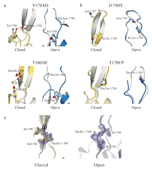
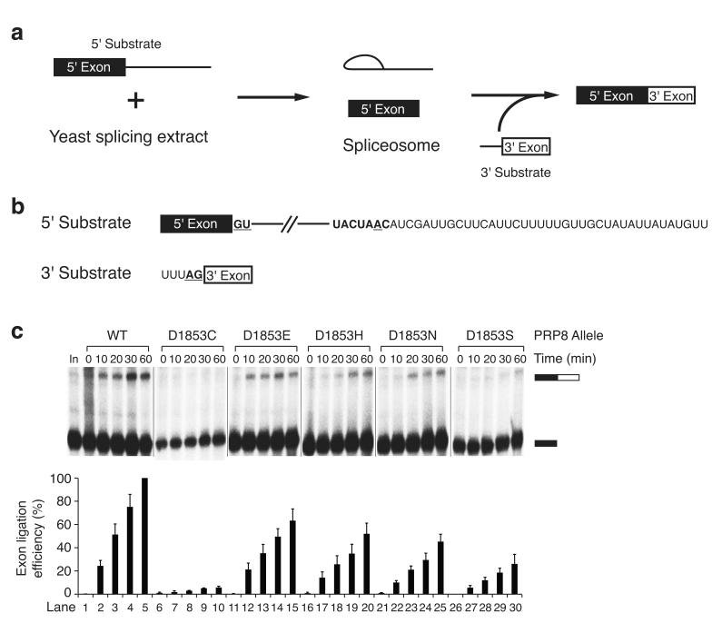
Splicing of pre-mRNAs in eukaryotes is catalyzed by the spliceosome, a large RNA-protein metalloenzyme. The catalytic center of the spliceosome involves a structure comprising the U2 and U6 snRNAs and includes a metal bound by U6 snRNA. The precise architecture of the splicesome active site, however, and the question of whether it includes protein components, remains unresolved. A wealth of evidence places the protein PRP8 at the heart of the spliceosome through assembly and catalysis. Here we provide evidence that the RNase H domain of PRP8 undergoes a conformational switch between the two steps of splicing, rationalizing yeast prp8 alleles that promote either the first or second step. We also show that this switch unmasks a metal-binding site involved in the second step. Together, these data establish that PRP8 is a metalloprotein that promotes exon ligation within the spliceosome.


Figures

Figure 1
Figure 1
Conformational switch in the PRP8 RH domain unmasks a Mg2+ binding site. (a) X-ray structure of the PRP8 RH domain. Superposition of the closed (yellow) and open (cyan) conformations observed in the asymmetric unit. (b) 2Fo-Fc maps contoured at 1.0σ showing octahedral coordination of Mg2+ (purple) bound in the open conformation of wild-type and R1865A PRP8 RH. (c) Detail of Mg2+ ion (purple) coordination by Asp1781 and inner-sphere waters (red) in the open conformation of PRP8 RH. (d) Superposition of the X-ray structure of the PRP8 RH domain closed (yellow) and open (cyan) conformations detailing the displacement of Thr1783 to allow Mg2+ coordination. The metal ion bound in the open conformation is not shown for clarity.
Figure 2
Figure 2
Characterization of PRP8 RH domain alleles. (a) Spot assays showing BSG ACT1-CUP1 reporter-dependent growth of yeast containing wild-type and mutant PRP8 alleles in the presence of the indicated concentration of Cu2+. (b) Denaturing PAGE analysis of RT-primer extension with 32P-labeled primer to examine steady-state splicing efficiencies in PRP8 mutant yeast strains. Primer extension of ACT1-CUP1 RNA with wild-type, BSC, or BSG sequences combined with PRP8 alleles. The products corresponding to the mRNA, pre-mRNA, and intron-lariat intermediate in the gel are indicated (top). Quantification of the first and second step efficiency is shown (bottom). BSG: branch site guanosine; BSC: branch site cytidine.
Figure 3
Figure 3
PRP8 mutant alleles favor distinct conformations within the PRP8 RH domain. Shown are superpositions of X-ray structures of wild-type (gray) with mutant closed (yellow) or mutant open (cyan) conformers. (a) (top) Detail showing formation of an additional hydrogen-bonding interaction to Tyr1786 in the closed conformation of the V1788D structure; (bottom) detail showing formation of a water (W) mediated additional hydrogen-bonding interaction to Tyr1786 in the closed conformation of the T1800E structure. (b) (top) Detail showing formation of a hydrogen bond across the loop with Asn1797 in the open conformation of the I1790Y structure; (bottom) detail showing disruption of the β-hairpin of the closed conformation in the T1789P structure. (c) X-ray structural analysis from a crystallographic mixing experiment with T1789P-R1865A and R1865A proteins. Shown are models for structures of the R1865A (gray) and T1789P-R1865A protein for the closed (yellow, left panel) and open (cyan, right panel) conformation. A 2Fo-Fc map contoured at 1σ calculated from the dataset of the T1789P-R1865A + R1865A mixture crystal is shown in dark blue.
Figure 4
Figure 4
PRP8 Asp1853 mutations selectively impair the second step of splicing. (a) Phenotypic analysis of PRP8 Asp1853 mutations. Serial dilutions of cells from strains harboring the indicated PRP8 alleles were spotted onto YPD medium; plates were incubated for 4 days at 16°C, 2 days at 30°C, or 3 days at 37°C and photographed. (b) Denaturing PAGE analysis of in vitro splicing of 32P-labeled actin pre-mRNA in extracts prepared from strains harboring the indicated Asp1853 PRP8 mutants (top). The positions of the mRNA, pre-mRNA, intron-lariat, and exon-lariat intermediate in the gel are indicated. Quantification of first and second step efficiencies is shown (bottom). (c) D1853C rescue by the second-step allele V1862Y. Growth of a dilution series of the D1853C-V1862Y yeast strain at 16°C, 30°C, and 37°C. Compare to growth of wild-type and D1853C strains in panel a. (d) Denaturing PAGE analysis comparing in vitro splicing of 32P-labeled actin pre-mRNA substrate in extracts prepared from wild-type, D1853C, and D1853C-V1862Y yeast strains.
Figure 5
Figure 5
Bimolecular exon ligation assay implicates PRP8 Asp 1853 in the second transesterification step of pre-mRNA splicing. (a) Schematic representation of bimolecular exon ligation reaction. (b) RNA substrates used in the yeast bimolecular exon ligation. (c) Bimolecular exon ligation assay for analysis of the effect of Asp1853 mutations on the second step of splicing. A 32P-labeled 5′ substrate RNA derived from ACT1 pre-mRNA was incubated under splicing conditions in wild-type and PRP8 Asp1853 mutant yeast extracts, chased with unlabeled RNA containing the 3′ splice site, and the products were analyzed by denaturing PAGE (top; Supplementary Fig. 5c). The positions of the 5′ exon and ligated product are indicated. Quantification of exon ligation efficiency is shown (bottom). Exon ligation efficiency was corrected for the contribution from a degradation product present in the input lane and was normalized to lane 5. Error bars indicate the standard deviation from three independent experiments.
Figure 6
Figure 6
PRP8 RH undergoes a conformational switch to present a functionally important metal ion to the spliceosome. (a) Structures of closed and open conformations of PRP8 RH. Residues corresponding to first and second step alleles used in this analysis are highlighted in blue, Mg2+ coordinating ligands in red, Mg2+ is purple, Thr1783 in yellow, and the peptide that crosslinks to the 5′ splice site in green. (b) Distinct conformational states of PRP8 are associated with the first and second transesterification steps of splicing. (c) Modeling of the PRP8 RH domain closed (yellow) and open (blue) conformations on the overall PRP8 structure (PDB ID 4I43) shown in gray. The Aar2 assembly factor structure has been removed for clarity. (d) The switch from closed (yellow) to open (blue) conformation positions a Mg2+ ion that promotes the second transesterification reaction in the active site of the spliceosome. PRP8 RH domain closed and open structures are modeled on the overall PRP8 structure shown in gray. The Mg2+ ion is highlighted in purple. Ends of a disordered loop that crosslinks to the branch region of the pre-mRNA before the second step are shown in green. (e) Aar2 sequesters the spliceosome active site cavity. The U5 snRNP assembly factor Aar2 (cyan) blocks the surface of the PRP8 RH domain (yellow) partially filling the active site cavity (amino acids 320–344, depicted as spheres) and stabilizes the PRP8 RH β-hairpin by extending the β-sheet (amino acids 345–353). Mg2+ coordinating residues in RH are shown in red.

Comment in

  • Toggling in the spliceosome.
    Abelson J. Abelson J. Nat Struct Mol Biol. 2013 Jun;20(6):645-7. doi: 10.1038/nsmb.2603. Nat Struct Mol Biol. 2013. PMID: 23739164 No abstract available.

References

    1. Jurica MS, Moore MJ. Pre-mRNA splicing: awash in a sea of proteins. Mol. Cell. 2003;12:5–14. - PubMed
    1. Rappsilber J, Ryder U, Lamond AI, Mann M. Large-scale proteomic analysis of the human spliceosome. Genome Res. 2002;12:1231–1245. - PMC - PubMed
    1. Zhou Z, Licklider LJ, Gygi SP, Reed R. Comprehensive proteomic analysis of the human spliceosome. Nature. 2002;419:182–185. - PubMed
    1. Madhani HD, Guthrie C. A novel base-pairing interaction between U2 and U6 snRNAs suggests a mechanism for the catalytic activation of the spliceosome. Cell. 1992;71:803–817. - PubMed
    1. Reyes JL, Gustafson EH, Luo HR, Moore MJ, Konarska MM. The C-terminal region of hPrp8 interacts with the conserved GU dinucleotide at the 5′ splice site. RNA. 1999;5:167–179. - PMC - PubMed

Publication types

MeSH terms

LinkOut - more resources