. 2013 Jun;33(11):2188-201.
doi: 10.1128/MCB.01637-12. Epub 2013 Mar 25.

Structural basis for activation of ZAP-70 by phosphorylation of the SH2-kinase linker

Affiliations

Structural basis for activation of ZAP-70 by phosphorylation of the SH2-kinase linker

Qingrong Yan et al. Mol Cell Biol. 2013 Jun.

Abstract

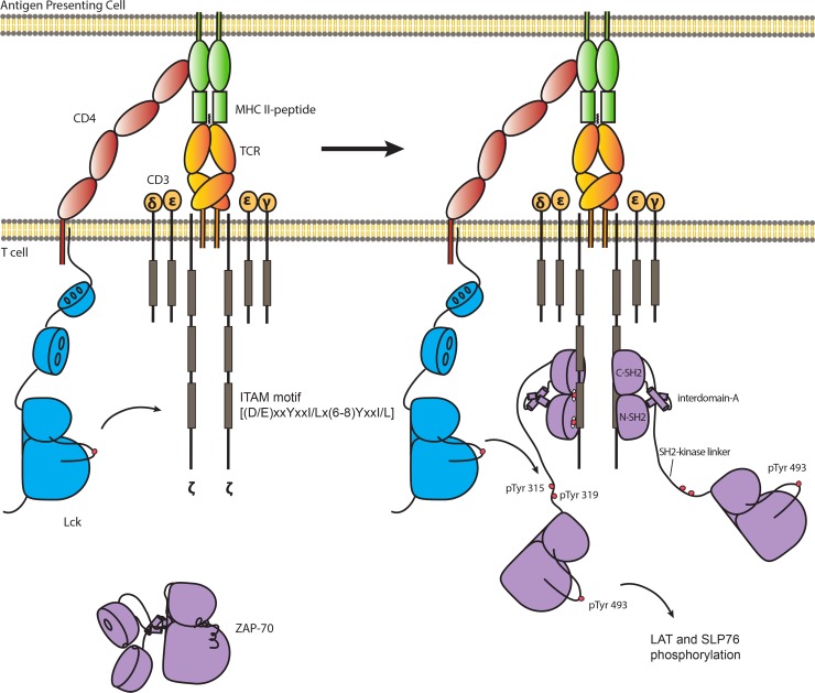
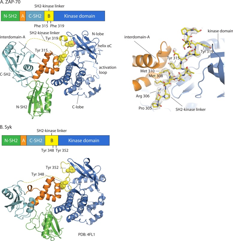
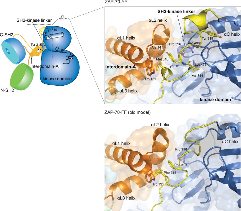
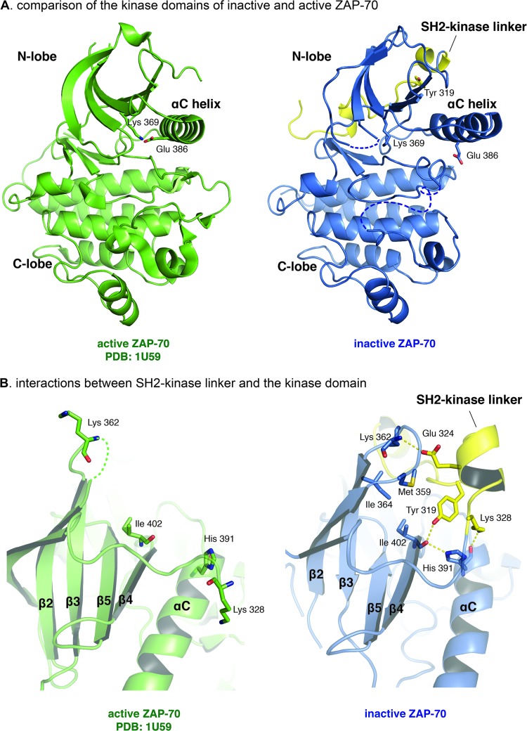
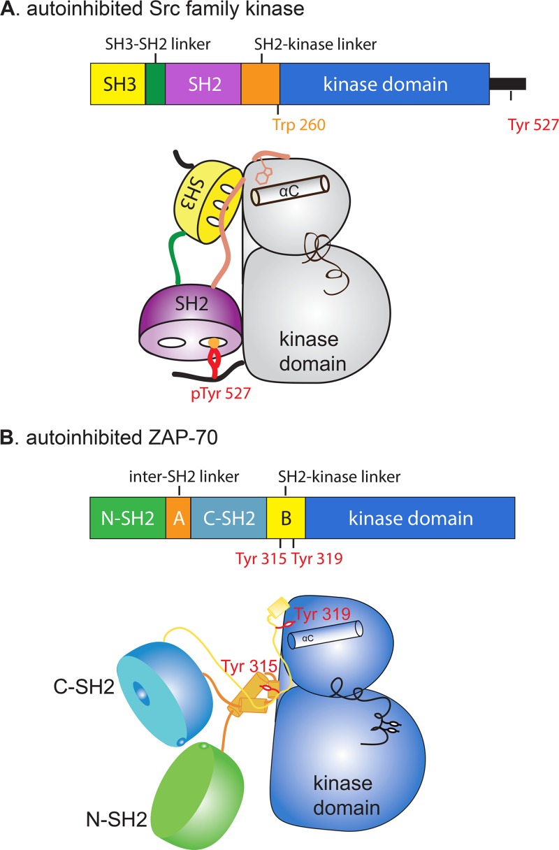
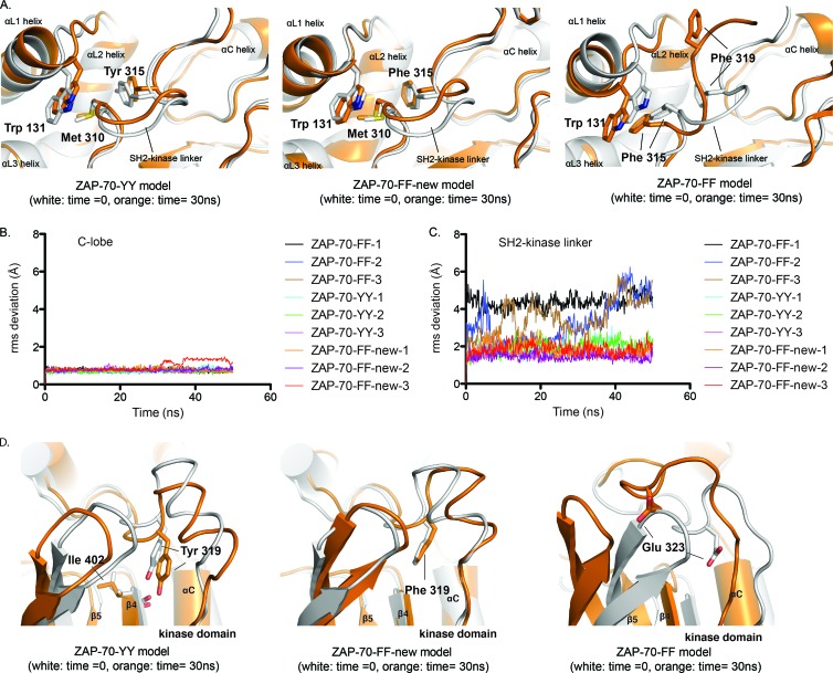
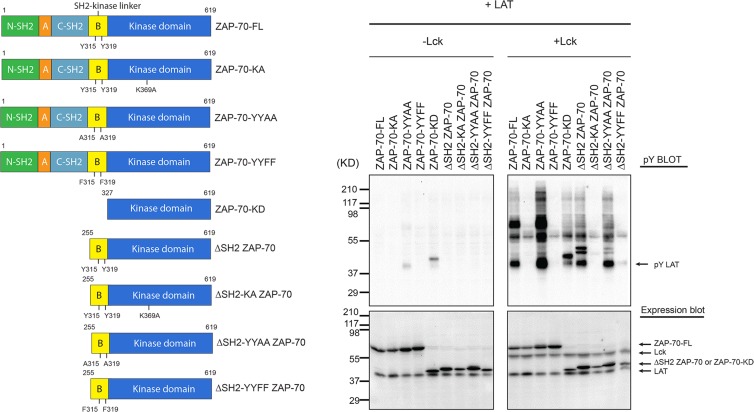
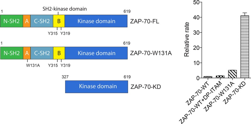
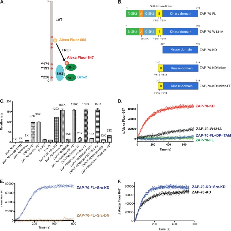
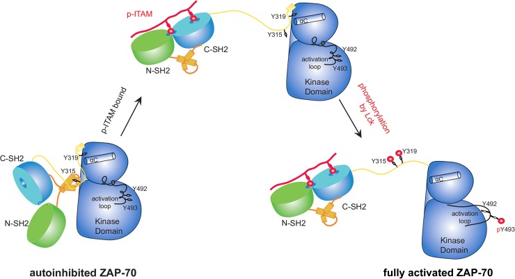
Serial activation of the tyrosine kinases Lck and ZAP-70 initiates signaling downstream of the T cell receptor. We previously reported the structure of an autoinhibited ZAP-70 variant in which two regulatory tyrosine residues (315 and 319) in the SH2-kinase linker were replaced by phenylalanine. We now present a crystal structure of ZAP-70 in which Tyr 315 and Tyr 319 are not mutated, leading to the recognition of a five-residue sequence register error in the SH2-kinase linker of the original crystallographic model. The revised model identifies distinct roles for these two tyrosines. As seen in a recently reported structure of the related tyrosine kinase Syk, Tyr 315 of ZAP-70 is part of a hydrophobic interface between the regulatory apparatus and the kinase domain, and the integrity of this interface would be lost upon engagement of doubly phosphorylated peptides by the SH2 domains. Tyr 319 is not necessarily dislodged by SH2 engagement, which activates ZAP-70 only ∼5-fold in vitro. In contrast, phosphorylation by Lck activates ZAP-70 ∼100-fold. This difference is due to the ability of Tyr 319 to suppress ZAP-70 activity even when the SH2 domains are dislodged from the kinase domain, providing stringent control of ZAP-70 activity downstream of Lck.


Figures

Fig 1
Fig 1
Phosphorylation of ZAP-70 is required to initiate T cell receptor signaling. TCR signaling is initiated by two tyrosine kinases: the Src family kinase Lck and ZAP-70. Activated Lck phosphorylates ITAMs in the intracellular tails of ζ chains of the TCR complex. Doubly phosphorylated ITAMs recruit ZAP-70 from the cytosol to the plasma membrane. Tyr 315, Tyr 319, and Tyr 493 of ZAP-70 are phosphorylated by Lck to fully activate ZAP-70, enabling ZAP-70 to phosphorylate two scaffold proteins, LAT and SLP-76, leading to the recruitment of effector proteins that stimulate T cell activation.
Fig 2
Fig 2
Overall structural organization of autoinhibited ZAP-70 and Syk. Domain organizations and crystal structures are shown for ZAP-70 (this work) (A) and Syk (PDB code 4FL2) (B). The two crystal structures are superimposed around the C lobes of the kinase domains. Disordered regions are depicted as dotted lines. The side chains of Tyr 315 and Tyr 319 in ZAP-70 and Tyr 348 and Tyr 352 in Syk are shown as spheres. A simulated-annealing composite omit map (A, right panel) (residues 305 to 323, contoured at 1.2 σ) is shown for the SH2-kinase linker of ZAP-70 (gray mesh representation).
Fig 3
Fig 3
Comparison of SH2-kinase linkers in the new, ZAP-70-YY model (top) and the original, ZAP-70-FF model (bottom).
Fig 4
Fig 4
Interactions between the SH2-kinase linker and the kinase domain stabilize the inactive conformation of the kinase domain. (A) Comparison of the kinase domains of inactive and active ZAP-70. (B) Interactions between the SH2-kinase linker and the kinase domain. (Left) Active ZAP-70 kinase domain (PDB code 1U59). (Right) Inactive ZAP-70 kinase domain with the SH2-kinase linker.
Fig 5
Fig 5
Cartoon representations of an autoinhibited Src family kinase and autoinhibited ZAP-70.
Fig 6
Fig 6
The conformation of the SH2-kinase linker in the ZAP-70-YY model is energetically favorable. (A) Overlays of instantaneous structures of the SH2-kinase linker at ∼30 ns from molecular dynamic trajectories obtained using the C lobe of the kinase domain as a reference for the alignment. (Left) ZAP-70-YY model. (Middle) ZAP-70-FF-new model. (Right) ZAP-70-FF model. (B) RMS deviations for C-α atoms in the C lobe of the kinase domain with respect to the initial positions. (C) RMS deviations for C-α atoms in the SH2-kinase linker with respect to the initial positions, using the C lobe of the kinase domain as a reference for the alignment. (D) Overlays of instantaneous structures of the N lobes of the kinase domains at ∼30 ns from molecular dynamic trajectories obtained using the C lobe of the kinase domain as a reference for the alignment. (Left) ZAP-70-YY model. (Middle) ZAP-70-FF-new model. (Right) ZAP-70-FF model.
Fig 7
Fig 7
In vivo kinase activity of ZAP-70 in HEK 293T cells. (Left) Domain architectures of various ZAP-70 truncation constructs. (Right) HEK 293 cells were transiently transfected with various constructs of ZAP-70 and LAT-FLAG together, with or without Lck, as indicated at the top. Cells were lysed in 2× SDS-PAGE sample buffer and analyzed by immunoblotting with an antibody against phosphotyrosine (monoclonal antibody 4G10). The expression levels of LAT-FLAG, ZAP-70, and Lck were assessed by probing with specific antibodies.
Fig 8
Fig 8
In vitro catalytic activity of ZAP-70 determined by coupled-kinase assay. (Left) Domain architectures of various ZAP-70 truncation constructs. (Right) Relative rates of phosphorylation of LAT by various ZAP-70 constructs.
Fig 9
Fig 9
Phosphorylation of ZAP-70 by Src family kinases fully activates ZAP-70 in vitro. (A) In vitro FRET-based approach to measure the catalytic activity of ZAP-70 for its physiological substrate, LAT. (B) Domain architectures of various ZAP-70 truncation constructs. (C) Relative rates of phosphorylation of LAT by ZAP-70 or Src family kinases. Src-KD, isolated c-Src kinase domain; Lck-FL, full-length Lck; DP-ITAM, doubly phosphorylated ITAM peptide. (D) Time course of phosphorylation of LAT as measured by the recruitment of Grb2. Red, isolated ZAP-70 kinase domain; black, ZAP-70-W131A; blue, full-length ZAP-70 bound to an excess amount of doubly phosphorylated ITAM peptide; green, full-length ZAP-70. (E) Time course of phosphorylation of LAT as measured by the recruitment of Grb2. Blue, full-length ZAP-70 plus Src kinase domain; brown, full-length ZAP-70 plus Src kinase domain with D386N mutation. (F) Time course of phosphorylation of LAT as measured by the recruitment of Grb2. Black, isolated ZAP-70 kinase domain; blue, isolated ZAP-70 kinase domain plus Src kinase domain.
Fig 10
Fig 10
Model of ZAP-70 activation by doubly phosphorylated ITAM peptide and Lck phosphorylation.

References

    1. Au-Yeung BB, Deindl S, Hsu LY, Palacios EH, Levin SE, Kuriyan J, Weiss A. 2009. The structure, regulation, and function of ZAP-70. Immunol. Rev. 228:41–57 - PubMed
    1. Sloan-Lancaster J, Presley J, Ellenberg J, Yamazaki T, Lippincott-Schwartz J, Samelson LE. 1998. ZAP-70 association with T cell receptor zeta (TCRzeta): fluorescence imaging of dynamic changes upon cellular stimulation. J. Cell Biol. 143:613–624 - PMC - PubMed
    1. Sloan-Lancaster J, Zhang W, Presley J, Williams BL, Abraham RT, Lippincott-Schwartz J, Samelson LE. 1997. Regulation of ZAP-70 intracellular localization: visualization with the green fluorescent protein. J. Exp. Med. 186:1713–1724 - PMC - PubMed
    1. Zhang W, Sloan-Lancaster J, Kitchen J, Trible RP, Samelson LE. 1998. LAT: the ZAP-70 tyrosine kinase substrate that links T cell receptor to cellular activation. Cell 92:83–92 - PubMed
    1. Bubeck Wardenburg J, Fu C, Jackman JK, Flotow H, Wilkinson SE, Williams DH, Johnson R, Kong G, Chan AC, Findell PR. 1996. Phosphorylation of SLP-76 by the ZAP-70 protein-tyrosine kinase is required for T-cell receptor function. J. Biol. Chem. 271:19641–19644 - PubMed

Publication types

MeSH terms

Associated data