. 2013;9(5):e1003390.
doi: 10.1371/journal.ppat.1003390. Epub 2013 May 23.

Structural and functional basis for inhibition of erythrocyte invasion by antibodies that target Plasmodium falciparum EBA-175

Affiliations

Structural and functional basis for inhibition of erythrocyte invasion by antibodies that target Plasmodium falciparum EBA-175

Edwin Chen et al. PLoS Pathog. 2013.

Abstract

Disrupting erythrocyte invasion by Plasmodium falciparum is an attractive approach to combat malaria. P. falciparum EBA-175 (PfEBA-175) engages the host receptor Glycophorin A (GpA) during invasion and is a leading vaccine candidate. Antibodies that recognize PfEBA-175 can prevent parasite growth, although not all antibodies are inhibitory. Here, using x-ray crystallography, small-angle x-ray scattering and functional studies, we report the structural basis and mechanism for inhibition by two PfEBA-175 antibodies. Structures of each antibody in complex with the PfEBA-175 receptor binding domain reveal that the most potent inhibitory antibody, R217, engages critical GpA binding residues and the proposed dimer interface of PfEBA-175. A second weakly inhibitory antibody, R218, binds to an asparagine-rich surface loop. We show that the epitopes identified by structural studies are critical for antibody binding. Together, the structural and mapping studies reveal distinct mechanisms of action, with R217 directly preventing receptor binding while R218 allows for receptor binding. Using a direct receptor binding assay we show R217 directly blocks GpA engagement while R218 does not. Our studies elaborate on the complex interaction between PfEBA-175 and GpA and highlight new approaches to targeting the molecular mechanism of P. falciparum invasion of erythrocytes. The results suggest studies aiming to improve the efficacy of blood-stage vaccines, either by selecting single or combining multiple parasite antigens, should assess the antibody response to defined inhibitory epitopes as well as the response to the whole protein antigen. Finally, this work demonstrates the importance of identifying inhibitory-epitopes and avoiding decoy-epitopes in antibody-based therapies, vaccines and diagnostics.


Conflict of interest statement

I have read the journal's policy and have the following conflicts: Coauthor B. Kim Lee Sim is the President and Chief Scientific Officer of Protein Potential. The authors from Washington University School of Medicine (Chen, Paing, Salinas and Tolia) have no affiliation to Protein Potential other than for pure interest in scientific collaboration. This does not alter any author's adherence to all the PLoS Pathogens' policies on sharing data and materials.

Figures

Figure 1
Figure 1. Crystal structure of RII/R217 Fab complex.
(A) Overall structure of the RII/R217 Fab complex shown in ribbon representation. The F1 domain of RII is colored in green, the F2 domain of RII is colored purple. The Fab heavy chain (VH) is in blue and the light chain (VL) in pink. The location of F2 β-finger is circled in black. (B) Ribbon representation of F2 mapping the R217 epitope. Residues contacted by the Fab are show in stick. Residues contacted by the heavy chain are colored blue, residues contacted by the Fab light chain are colored pink, and residues contacted by both chains are in beige. Residues not contacted by the antibody are in purple. (C) Surface representation of F2 mapping the R217 epitope. Color scheme as in B. (D) Surface representation of the R217 Fab, mapping heavy chain residues (blue) that contact F2 (purple). The light chain is shown in white. (E) Surface representation of the R217 Fab, mapping light chain residues (pink) that contact F2 (purple). The heavy chain is shown in white.
Figure 2
Figure 2. SAXS analysis of RII/R217 Fab.
(A) Plot of scattering intensity (I) against scattering momentum (Q) and statistical fit of theoretical scatter from the RII/R217 Fab crystal structure (red line) with experimental SAXS profile (black). (B) Overlay of ab initio averaged reconstruction model of SAXS data with crystal structure. RII F1 domain is green, RII F2 domain is purple, the Fab heavy chain (VH) is blue and the Fab light chain (VL) is pink. The ab initio envelope is colored grey.
Figure 3
Figure 3. Crystal structure of F1/R218 Fab complex.
(A) Overall structure of the F1/R218 Fab complex shown in ribbon representation. The F2 domain of RII is colored in green. The Fab heavy chain (VH) is in blue and the light chain (VL) in pink. (B) Ribbon representation of F1 mapping the R218 epitope. Residues contacted by the Fab are show in stick. Residues contacted by the heavy chain are colored blue, residues contacted by the Fab light chain are colored orange, and residues contacted by both chains are in beige. Residues not contacted by the antibody are in green. (C) Surface representation of F2 mapping the R217 epitope. Color scheme as in B. (D) Surface representation of the R218 Fab, mapping heavy chain residues (blue) that contact F2 (green). The light chain is shown in white. (E) Surface representation of the R218 Fab, mapping light chain residues (orange) that contact F2 (green). The heavy chain is shown in white.
Figure 4
Figure 4. Antibody epitope identification by immunofluorescence assay.
DBL domain proteins were surface expressed on HEK293 cells and probed with R217 or R218 as primary and Alexafluor-546 labeled anti-IgG1 as secondary. Green channel shows GFP tagged expressed protein. Red channel shows Alexafluor-546 labeled proteins on HEK293 cell surface. Merged channel shows overlap between green and red channels.
Figure 5
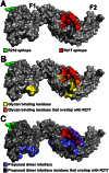
Figure 5. The R217 epitope overlaps with glycan binding residues and the proposed dimer interface, while the R218 epitope is far removed from these regions.
(A) Surface representation of RII (grey) with the R217 (red) and R218 (green) epitopes. (B) The R217 epitope overlaps with glycan binding residues, including K341, N417 and R422 with demonstrated roles in erythrocyte binding . The R218 epitope is located on the opposite face of RII away from glycan binding residues. Glycan binding residues are in yellow, glycan binding residues that overlap with the R217 epitope are in orange, the R217 epitope is in red, the R218 epitope is in green and RII is in grey. (C) The R217 epitope overlaps with proposed dimer interface residues while the R218 epitope is far removed from the proposed dimer interface. Proposed dimer interface residues are in blue, proposed dimer interface residues that overlap with the R217 epitope are in purple, the R217 epitope is in red, the R218 epitope is in green and RII is in grey.
Figure 6
Figure 6. Direct antibody-inhibition of GpA binding by RII.
(A) RII binds to GpA (lane 1), but not to neuraminidase treated GpA (GpA-NA – lane 2). Addition of R217 IgG or Fab prevents RII from binding GpA (lane 3 and 4). Neither R218 IgG (lane 5), R218 Fab (lane 6), control IgG (lane 7) nor control Fab (lane 8), block RII/GpA receptor binding. Binding is specific as neuraminidase treatment prevents binding in all cases (lanes 2 and 9–14). Note that an increased signal is observed with R218 IgG over R218 Fab (compare lanes 5 and 6) suggesting bivalent binding. This assay was performed with RII at 3 µM and antibody or Fab fragments at 6 µM. (B) Titration of R217 demonstrates the specificity of interaction as all concentrations around and above the available RII concentration (3 µM) prevents binding (lanes 6–10). At concentrations below the available RII binding occurs as not all the RII is bound by R217 (lanes 3–5). (C) Titration of R218 demonstrates that R218 is unable to directly prevent GpA binding at any concentration.

References

    1. Orlandi PA, Klotz FW, Haynes JD (1992) A malaria invasion receptor, the 175-kilodalton erythrocyte binding antigen of Plasmodium falciparum recognizes the terminal Neu5Ac(alpha 2–3)Gal- sequences of glycophorin A. J Cell Biol 116: 901–909. - PMC - PubMed
    1. Adams JH, Sim BK, Dolan SA, Fang X, Kaslow DC, et al. (1992) A family of erythrocyte binding proteins of malaria parasites. Proc Natl Acad Sci U S A 89: 7085–7089. - PMC - PubMed
    1. Camus D, Hadley TJ (1985) A Plasmodium falciparum antigen that binds to host erythrocytes and merozoites. Science 230: 553–556. - PubMed
    1. Sim BK, Chitnis CE, Wasniowska K, Hadley TJ, Miller LH (1994) Receptor and ligand domains for invasion of erythrocytes by Plasmodium falciparum. Science 264: 1941–1944. - PubMed
    1. Sim BK, Orlandi PA, Haynes JD, Klotz FW, Carter JM, et al. (1990) Primary structure of the 175K Plasmodium falciparum erythrocyte binding antigen and identification of a peptide which elicits antibodies that inhibit malaria merozoite invasion. J Cell Biol 111: 1877–1884. - PMC - PubMed

Publication types

MeSH terms